MAB3286, Mouse IgG
2Bisotype), anti-HSP60 (Catalog #
MAB1800, Mouse IgG
2Bisotype), anti-GAPDH (Catalog #
MAB5718,Mouse IgG
1isotype), anti-Peroxiredoxin 6 (Catalog #
MAB3490, Mouse IgG
3isotype), and anti-SOD1 (Catalog #
MAB3418, Mouse IgG
2Aisotype). The left panel was then probed with 0.5 µg/mL of Rabbit Anti-Mouse IgG
3(Catalog # MAB9675) followed by HRP-conjugated Anti-Rabbit IgG Secondary Antibody (Catalog #
HAF008). A specific bands was detected for IgG
3isotype antibody at approximately 26 kDa (as indicated). The right panel was probed with HRP-conjugated Anti-Mouse IgG Secondary Antibody (Catalog #
HAF018) but not Rabbit Anti-Mouse IgG
2BMonoclonal Antibody (Catalog # MAB9675). This experiment was conducted under reducing conditions and using
Immunoblot Buffer Group 1." class="big_lightbox">
MAB3286, Mouse IgG
2Bisotype), anti-HSP60 (Catalog #
MAB1800, Mouse IgG
2Bisotype), anti-GAPDH (Catalog #
MAB5718,Mouse IgG
1isotype), anti-Peroxiredoxin 6 (Catalog #
MAB3490, Mouse IgG
3isotype), and anti-SOD1 (Catalog #
MAB3418, Mouse IgG
2Aisotype). The left panel was then probed with 0.5 µg/mL of Rabbit Anti-Mouse IgG
3(Catalog # MAB9675) followed by HRP-conjugated Anti-Rabbit IgG Secondary Antibody (Catalog #
HAF008). A specific bands was detected for IgG
3isotype antibody at approximately 26 kDa (as indicated). The right panel was probed with HRP-conjugated Anti-Mouse IgG Secondary Antibody (Catalog #
HAF018) but not Rabbit Anti-Mouse IgG
2BMonoclonal Antibody (Catalog # MAB9675). This experiment was conducted under reducing conditions and using
Immunoblot Buffer Group 1." alt="Western blot shows lysates of HepG2 and Jurkat cell lines. PVDF membrane was probed with anti-HSP90 (Catalog #
MAB3286, Mouse IgG
2Bisotype), anti-HSP60 (Catalog #
MAB1800, Mouse IgG
2Bisotype), anti-GAPDH (Catalog #
MAB5718,Mouse IgG
1isotype), anti-Peroxiredoxin 6 (Catalog #
MAB3490, Mouse IgG
3isotype), and anti-SOD1 (Catalog #
MAB3418, Mouse IgG
2Aisotype). The left panel was then probed with 0.5 µg/mL of Rabbit Anti-Mouse IgG
3(Catalog # MAB9675) followed by HRP-conjugated Anti-Rabbit IgG Secondary Antibody (Catalog #
HAF008). A specific bands was detected for IgG
3isotype antibody at approximately 26 kDa (as indicated). The right panel was probed with HRP-conjugated Anti-Mouse IgG Secondary Antibody (Catalog #
HAF018) but not Rabbit Anti-Mouse IgG
2BMonoclonal Antibody (Catalog # MAB9675). This experiment was conducted under reducing conditions and using
Immunoblot Buffer Group 1." class="big_thumb" />
MAB9186, Mouse IgG
3isotype), followed by Rabbit anti-Mouse IgG
3-specific secondary antibody (Catalog # MAB9675, filled histogram), Rabbit anti-Mouse IgG
1-specific (Catalog #
MAB9627, blue line), Rabit anti-Mouse IgG
2A-specific (Catalog #
MAB0033, red line), or Rabbit anti-Mouse IgG
2B-specific (Catalog #
MAB9636, black line) secondary antibody, then PE-conjugated Goat anti-Rabbit secondary antibody (Catalog #
F0110). View our protocol for
Staining Membrane-associated Proteins." class="big_lightbox">
MAB9186, Mouse IgG
3isotype), followed by Rabbit anti-Mouse IgG
3-specific secondary antibody (Catalog # MAB9675, filled histogram), Rabbit anti-Mouse IgG
1-specific (Catalog #
MAB9627, blue line), Rabit anti-Mouse IgG
2A-specific (Catalog #
MAB0033, red line), or Rabbit anti-Mouse IgG
2B-specific (Catalog #
MAB9636, black line) secondary antibody, then PE-conjugated Goat anti-Rabbit secondary antibody (Catalog #
F0110). View our protocol for
Staining Membrane-associated Proteins." class="big_thumb" />
 MAB1435, filled histogram) or Mouse IgG Isotype Control Antibody (Catalog # MAB007, open histogram), followed by Phycoerythrin-conjugated Anti-Mouse IgG Secondary Antibody (Catalog # F0102B)." alt="NTera-2 human testicular embryonic carcinoma cell line was stained with Mouse Anti-Human/Mouse SSEA-4 Monoclonal Antibody (Catalog # MAB1435, filled histogram) or Mouse IgG Isotype Control Antibody (Catalog # MAB007, open histogram), followed by Phycoerythrin-conjugated Anti-Mouse IgG Secondary Antibody (Catalog # F0102B)." class="big_thumb" />
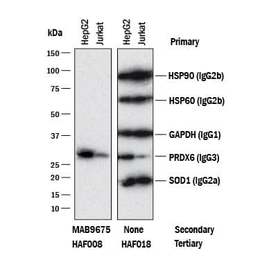
 MAB3286, Mouse IgG2Bisotype), anti-HSP60 (Catalog # MAB1800, Mouse IgG2Bisotype), anti-GAPDH (Catalog # MAB5718,Mouse IgG1isotype), anti-Peroxiredoxin 6 (Catalog # MAB3490, Mouse IgG3isotype), and anti-SOD1 (Catalog # MAB3418, Mouse IgG2Aisotype). The left panel was then probed with 0.5 µg/mL of Rabbit Anti-Mouse IgG3(Catalog # MAB9675) followed by HRP-conjugated Anti-Rabbit IgG Secondary Antibody (Catalog # HAF008). A specific bands was detected for IgG3isotype antibody at approximately 26 kDa (as indicated). The right panel was probed with HRP-conjugated Anti-Mouse IgG Secondary Antibody (Catalog # HAF018) but not Rabbit Anti-Mouse IgG2BMonoclonal Antibody (Catalog # MAB9675). This experiment was conducted under reducing conditions and using Immunoblot Buffer Group 1." alt="Western blot shows lysates of HepG2 and Jurkat cell lines. PVDF membrane was probed with anti-HSP90 (Catalog # MAB3286, Mouse IgG2Bisotype), anti-HSP60 (Catalog # MAB1800, Mouse IgG2Bisotype), anti-GAPDH (Catalog # MAB5718,Mouse IgG1isotype), anti-Peroxiredoxin 6 (Catalog # MAB3490, Mouse IgG3isotype), and anti-SOD1 (Catalog # MAB3418, Mouse IgG2Aisotype). The left panel was then probed with 0.5 µg/mL of Rabbit Anti-Mouse IgG3(Catalog # MAB9675) followed by HRP-conjugated Anti-Rabbit IgG Secondary Antibody (Catalog # HAF008). A specific bands was detected for IgG3isotype antibody at approximately 26 kDa (as indicated). The right panel was probed with HRP-conjugated Anti-Mouse IgG Secondary Antibody (Catalog # HAF018) but not Rabbit Anti-Mouse IgG2BMonoclonal Antibody (Catalog # MAB9675). This experiment was conducted under reducing conditions and using Immunoblot Buffer Group 1." class="big_thumb" />
 HAF018). A specific band was detected for IgG3at approximately 240 kDa (as indicated). Human IgG (Catalog # MAB1101) is shown as a loading control.This experiment was conducted under non-reducing conditions and using Immunoblot Buffer Group 1." alt="Western blot shows lysates of purified human IgG1(negative control), purified human IgG2(negative control), purified human IgG3, and human purified human IgG4(negative control). PVDF membrane was probed with 2 µg/mL of Mouse Anti-Human IgG3Monoclonal Antibody (Catalog # MAB10122) followed by HRP-conjugated Anti-Mouse IgG Secondary Antibody (Catalog # HAF018). A specific band was detected for IgG3at approximately 240 kDa (as indicated). Human IgG (Catalog # MAB1101) is shown as a loading control.This experiment was conducted under non-reducing conditions and using Immunoblot Buffer Group 1." class="big_thumb" />


![Western Blot: Mouse IgG3 Isotype Control (MG3) [NBP1-96777] - SDS-PAGE of Mouse IgG3. Lane 1: Non-Reduced Human IgG. Lane 2: 5uL OPAL Pre-stained Marker MB-210-0500. Lane 3: Reduced Mouse IgG3. Load: 1ug per lane. Predicted/Observed size: Non-reduced at 180-245 kDa, Reduced at 55, 25 kDa.](http://images.novusbio.com/fullsize/Mouse-IgG3-Isotype-Control-MG3-Western-Blot-NBP1-96777-img0003.jpg "Western Blot: Mouse IgG3 Isotype Control (MG3) [NBP1-96777] - SDS-PAGE of Mouse IgG3. Lane 1: Non-Reduced Human IgG. Lane 2: 5uL OPAL Pre-stained Marker MB-210-0500. Lane 3: Reduced Mouse IgG3. Load: 1ug per lane. Predicted/Observed size: Non-reduced at 180-245 kDa, Reduced at 55, 25 kDa.")
![Dot Blot: Mouse IgG3 Isotype Control (MG3) [NBP1-96777] - A three-fold serial dilution of Fluorescein conjugated Mouse IgG3 subclass starting at 200ug was spotted onto 0.45 um nitrocellulose. Image from the FITC version of this antibody.](http://images.novusbio.com/fullsize/Mouse-IgG3-Isotype-Control-MG3-Dot-Blot-NBP1-96777-img0002.jpg)
![SDS-PAGE of Mouse IgG3 Isotype Control (MG3). Lane 1: Non-Reduced Human IgG. Lane 2: [5µL] Opal Pre-stained Marker . Lane 3: Reduced Mouse IgG3 Isotype Control (MG3). Load: 1µg per lane. Predicted/Observed size: Non-reduced at 180-245 kDa, Reduced at 55, 25 kDa.](http://images.novusbio.com/fullsize/nbp1-96777_mouse-mouse-igg3-isotype-control-mg3-1872025818710.jpg)
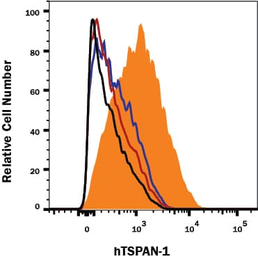
 MAB9186, Mouse IgG3isotype), followed by Rabbit anti-Mouse IgG3-specific secondary antibody (Catalog # MAB9675, filled histogram), Rabbit anti-Mouse IgG1-specific (Catalog #
 ![Immunohistochemistry: IgG3 Antibody (RM119) [Unconjugated] [NBP2-62125] - Immunohistochemistry of Human Lymphoid Tissue using NBP2-62125.](http://images.novusbio.com/fullsize/IgG3-Antibody-RM119-Unconjugated-Immunohistochemistry-NBP2-62125-img0003.jpg "Immunohistochemistry: IgG3 Antibody (RM119) [Unconjugated] [NBP2-62125] - Immunohistochemistry of Human Lymphoid Tissue using NBP2-62125.")
![ELISA: IgG3 Antibody (RM119) - Azide and BSA Free [NBP2-62125] - ELISA of human immunoglobulins shows RM119 only reacts to human IgG3. No cross reactivity with other human immunoglobulins, mouse, rat, or goat IgG.](http://images.novusbio.com/fullsize/nbp2-62125_rabbit-monoclonal-igg3-antibody-rm119-elisa-18920252340352.png)
![ELISA: IgG3 Antibody (RM119) - Azide and BSA Free [NBP2-62125] - A titer ELISA using RM119. The plate was coated with different amounts of human IgG3. A serial dilution of RM119 was used as the primary antibody.](http://images.novusbio.com/fullsize/nbp2-62125_rabbit-monoclonal-igg3-antibody-rm119-elisa-18920252341547.png)
![ELISA: Mouse IgG3 ELISA Kit (Colorimetric) [NBP2-68033] - Samples were spiked with high concentrations of Mouse IgG3 and diluted with Reference Standard & Sample Diluent to produce samples with values within the range of the assay.](http://images.novusbio.com/fullsize/nbp2-68033_mouse-igg3-elisa-kit-colorimetric-132202416204038.jpg "ELISA: Mouse IgG3 ELISA Kit (Colorimetric) [NBP2-68033] - Samples were spiked with high concentrations of Mouse IgG3 and diluted with Reference Standard & Sample Diluent to produce samples with values within the range of the assay.")
![ELISA: Mouse IgG3 ELISA Kit (Colorimetric) [NBP2-68033] - Standard Curve Reference](http://images.novusbio.com/fullsize/nbp2-68033_mouse-igg3-elisa-kit-colorimetric-432024903528.jpg)
![Western Blot: IgG3 Antibody (6Q4J8) [NBP3-15876] - Western blot analysis of extracts of human plasma, using IgG3 antibody (NBP3-15876) at 1:3000 dilution. Secondary antibody: HRP Goat Anti-Rabbit IgG (H+L) at 1:10000 dilution. Lysates/proteins: 25ug per lane. Blocking buffer: 3% nonfat dry milk in TBST. Detection: ECL Basic Kit. Exposure time: 1s.](http://images.novusbio.com/fullsize/IgG3-Antibody-6Q4J8-Western-Blot-NBP3-15876-img0001.jpg "Western Blot: IgG3 Antibody (6Q4J8) [NBP3-15876] - Western blot analysis of extracts of human plasma, using IgG3 antibody (NBP3-15876) at 1:3000 dilution. Secondary antibody: HRP Goat Anti-Rabbit IgG (H+L) at 1:10000 dilution. Lysates/proteins: 25ug per lane. Blocking buffer: 3% nonfat dry milk in TBST. Detection: ECL Basic Kit. Exposure time: 1s.")
![ELISA: IgG3 Antibody (RM218) - Azide and BSA Free [NBP2-62010] - ELISA of mouse immunoglobulins shows RM218 reacts to both mouse IgG3 kappa and IgG3 lambda; No cross reactivity with mouse IgG1, IgG2a, IgG2b, IgM, IgA, IgE, human IgG, or rat IgG. The plate was coated with 50 ng/well of different immunoglobulins. 200 ng/mL, 50 ng/mL, or 10 ng/mL of RM218 was used as the primary antibody. An alkaline phosphatase conjugated anti-rabbit IgG as the secondary antibody.](http://images.novusbio.com/fullsize/nbp2-62010_rabbit-monoclonal-igg3-antibody-rm218-elisa-189202523474816.png "ELISA: IgG3 Antibody (RM218) - Azide and BSA Free [NBP2-62010] - ELISA of mouse immunoglobulins shows RM218 reacts to both mouse IgG3 kappa and IgG3 lambda; No cross reactivity with mouse IgG1, IgG2a, IgG2b, IgM, IgA, IgE, human IgG, or rat IgG. The plate was coated with 50 ng/well of different immunoglobulins. 200 ng/mL, 50 ng/mL, or 10 ng/mL of RM218 was used as the primary antibody. An alkaline phosphatase conjugated anti-rabbit IgG as the secondary antibody.")
![ELISA: IgG3 Antibody (RM218) - Azide and BSA Free [NBP2-62010] - A titer ELISA of mouse IgG3. The plate was coated with different amounts of mouse IgG3. A serial dilution of RM218 was used as the primary antibody. An alkaline phosphatase conjugated anti-rabbit IgG as the secondary antibody.](http://images.novusbio.com/fullsize/nbp2-62010_rabbit-monoclonal-igg3-antibody-rm218-elisa-189202523394323.png)


