After August 17, 2026, Novus Biologicals products and services will no longer be available on this website; you will access all products and services on rndsystems.com. Create your R&D Systems online account today.
HAF016). A specific band was detected for CKAP4/p63 at approximately 63 kDa (as indicated). This experiment was conducted under reducing conditions and using Immunoblot Buffer Group 8." class="big_lightbox" target="_blank">
NL012). Cytokeratin 14 was also detected using Mouse Anti-Human Cytokeratin 14 Monoclonal Antibody (MAB3164) and stained using the NorthernLights™ 557-conjugated Anti-Mouse IgG Secondary Antibody (red; NL007). Cells were counterstained with DAPI (blue). Specific staining of CKAP4/p63 was localized to perinuclear space and cell membrane. View our protocol for Fluorescent ICC Staining of Cells on Coverslips." class="big_lightbox" target="_blank">
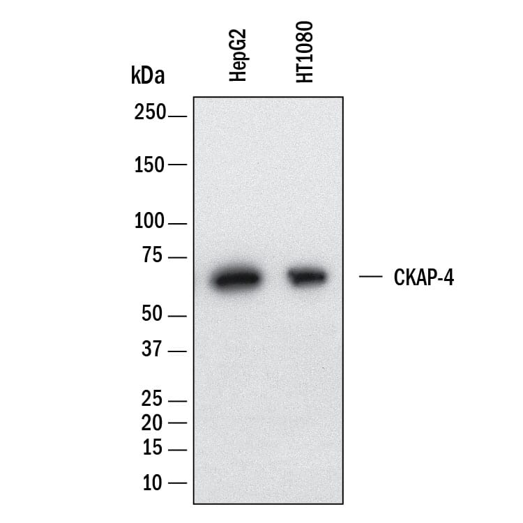
HAF018). A specific band was detected for CKAP4/p63 at approximately 65 kDa (as indicated). This experiment was conducted under reducing conditions and using Western Blot Buffer Group 1." class="big_lightbox" target="_blank">
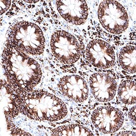
VC001). Before incubation with the primary antibody, tissue was subjected to heat-induced epitope retrieval using VisUCyte Antigen Retrieval Reagent-Basic (Catalog # VCTS021). Tissue was stained using DAB (brown) and counterstained with hematoxylin (blue). Specific staining was localized to the cell surface and cytoplasm. View our protocol for IHC Staining with VisUCyte HRP Polymer Detection Reagents." class="big_lightbox" target="_blank">
VC001). Before incubation with the primary antibody, tissue was subjected to heat-induced epitope retrieval using VisUCyte Antigen Retrieval Reagent-Basic (Catalog # VCTS021). Tissue was stained using DAB (brown) and counterstained with hematoxylin (blue). Specific staining was localized to the cell surface and cytoplasm. View our protocol for IHC Staining with VisUCyte HRP Polymer Detection Reagents." class="big_lightbox" target="_blank">
VC001). Before incubation with the primary antibody, tissue was subjected to heat-induced epitope retrieval using VisUCyte Antigen Retrieval Reagent-Basic (Catalog # VCTS021). Tissue was stained using DAB (brown) and counterstained with hematoxylin (blue). Specific staining was localized to the cell surface and cytoplasm. View our protocol for IHC Staining with VisUCyte HRP Polymer Detection Reagents." class="big_lightbox" target="_blank">
NL007) and counterstained with DAPI (blue). Specific staining was localized to the endoplasmic reticulum membrane. View our protocol for Fluorescent ICC Staining of Cells on Coverslips." class="big_lightbox" target="_blank">
5897-DK) is immobilized at 1 µg/mL (100 µL/well), Recombinant Mouse CKAP4/p63 binds with an ED50 of 35-280 ng/mL." class="big_lightbox" target="_blank">
Cytoskeleton-associated protein 4 (CKAP4) is also known as p63 and cytoskeleton-linking membrane protein 63kDa (CLIMP-63). It was first described as a palmitoylated single-pass transmembrane receptor that localized to the rough endoplasmic reticulum as well as the cell membrane. CKAP4 has been shown to help anchor endoplasmic reticulum to microtubules.
![Western Blot CKAP4/p63 Antibody [Unconjugated]](http://images.novusbio.com/images/antibody/CKAP4_AF7355_Western_Blot_11997.jpg)
![Western Blot CKAP4/p63 Antibody (1086526) [Unconjugated]](http://images.novusbio.com/images/antibody/mab11600_human-ckap4-p63-mab-clone-1086526-western-blot-2692024142928..jpg)

![Binding Activity CKAP4/p63 [Unconjugated]](http://images.novusbio.com/images/protein/CKAP4_9734CK_1884.jpg)

![Immunocytochemistry CKAP4/p63 Antibody [Unconjugated]](http://images.novusbio.com/images/antibody/CKAP4_AF7355_Immunocytochemistry_12002.jpg)
![Immunocytochemistry CKAP4/p63 Antibody [Unconjugated]](http://images.novusbio.com/images/antibody/CKAP4_AF7355_Immunocytochemistry__Immunofluorescence_17000.jpg)
![Simple Western CKAP4/p63 Antibody [Unconjugated]](http://images.novusbio.com/fullsize/antibody/af7355_human-ckap4-p63-affinity-purified-polyclonal-ab-simple-western-157202493849.jpg)
![Immunohistochemistry CKAP4/p63 Antibody (1086526) [Unconjugated]](http://images.novusbio.com/images/antibody/mab11600_human-ckap4-p63-mab-clone-1086526-immunohistochemistry-259202495646..jpg)
![Immunohistochemistry CKAP4/p63 Antibody (1086526) [Unconjugated]](http://images.novusbio.com/images/antibody/mab11600_human-ckap4-p63-mab-clone-1086526-immunohistochemistry-25920249588..jpg)
![Immunohistochemistry CKAP4/p63 Antibody (1086526) [Unconjugated]](http://images.novusbio.com/images/antibody/mab11600_human-ckap4-p63-mab-clone-1086526-immunohistochemistry-259202495918..jpg)
![Immunocytochemistry CKAP4/p63 Antibody (1086526) [Unconjugated]](http://images.novusbio.com/images/antibody/mab11600_human-ckap4-p63-mab-clone-1086526-immunocytochemistry-259202410422..jpg)
![Simple Western CKAP4/p63 Antibody (1086526) [Unconjugated]](http://images.novusbio.com/fullsize/antibody/mab11600_human-ckap4-p63-mab-clone-1086526-simple-western-2692024143221.jpg)