
| Reactivity | All-NASpecies Glossary |
| Applications | WB, Flow, ICC/IF, IHC, IP, KO, Single-Cell Western |
| Clone | 1C51 |
| Clonality | Monoclonal |
| Host | Mouse |
| Conjugate | Unconjugated |
| Format | BSA Free |
| Concentration | 1 mg/ml |
| Immunogen | This mCherry Antibody (1C51) was developed against recombinant full-length mCherry purified from E. coli. |
| Specificity | This mCherry Antibody (1C51) does not cross react with GFP. |
| Isotype | IgG2a |
| Clonality | Monoclonal |
| Host | Mouse |
| Purity | Protein G purified |
| Innovator's Reward | Test in a species/application not listed above to receive a full credit towards a future purchase. |
| Dilutions |
|
|
| Application Notes | Use in Flow reported in scientific literature (PMID:33335127). Use in IHC and IHC-P reported in scientific literature (PMID: 27396338 and 27716840 respectively). mCherry antibody validated for IHC-Frozen from a verified customer review. Use in Immunoprecipitation reported in scientific literature (PMID: 33008892). Use in Knockout Validation was reported in scientific literature (PMID: 32547960). |
|
| Theoretical MW | 27 kDa. Disclaimer note: The observed molecular weight of the protein may vary from the listed predicted molecular weight due to post translational modifications, post translation cleavages, relative charges, and other experimental factors. |
|
| Reviewed Applications |
|
|
| Publications |
|
| Storage | Store at 4C short term. Aliquot and store at -20C long term. Avoid freeze-thaw cycles. |
| Buffer | 50% PBS, 50% glycerol |
| Preservative | 0.035% Sodium Azide |
| Concentration | 1 mg/ml |
| Purity | Protein G purified |
| Images | Ratings | Applications | Species | Date | Details | ||||||||||
|---|---|---|---|---|---|---|---|---|---|---|---|---|---|---|---|
Enlarge |
reviewed by:
Verified Customer |
WB | Human | 04/19/2019 |
Summary
Comments
|
||||||||||
Enlarge |
reviewed by:
Verified Customer |
Immunofluorescence - fixed-frozen | Mouse | 10/21/2018 |
Summary
Comments
|
||||||||||
|
reviewed by:
Verified Customer |
WB | Human | 03/17/2017 |
Summary
|
Secondary Antibodies |
Isotype Controls |
Research Areas for mCherry Antibody (NBP1-96752)Find related products by research area.
|
|
Successful Transplantation of Friedreich Ataxia Induced Pluripotent Stem Cell (iPSC)-Derived Sensory Neurons in Dorsal Root Ganglia of Adult Rodents Jamshed Arslan, Pharm D, PhD The dorsal root ganglia (DRG) are a collection of cell bodies of sensory nerves carrying sensory information – including nociception, mechanoreception and proprioception – from periphera... Read full blog post. |
|
Autophagy and RAS signaling: Clinical implications By Christina Towers, PhD The cellular recycling process known as autophagy is currently being targeted in over 60 clinical trials focused on treating different types of cancer1. To date, the only autophagy-targeted ... Read full blog post. |
|
Read full blog post. |
|
Read full blog post. |
|
How to visualize autophagy by microscopy By Christina Towers, PhD Autophagy is a recycling process that relies on the formation of a unique organelle termed an autophagosome. An elegant way to monitor autophagy is through various microscopy techniques to... Read full blog post. |
|
Read full blog post. |
|
Animal Models to Study Autophagy By Christina Towers, PhD What is autophagy?Autophagy is the catabolic process that degrades cytoplasmic material via the lysosome. The process of macroautophagy was originally characterized in yeast, where the... Read full blog post. |
|
Application Focus: I see an increase in LC3, now what? By Christina Towers, PhD. Autophagy is highly conserved and tightly regulated process that all cell types use to recycle nutrients, particularly in the instance of stress1. As a result, even sm... Read full blog post. |
The concentration calculator allows you to quickly calculate the volume, mass or concentration of your vial. Simply enter your mass, volume, or concentration values for your reagent and the calculator will determine the rest.
5 | |
4 | |
3 | |
2 | |
1 |
| Verified Customer 04/19/2019 |
||
| Application: | WB | |
| Species: | Human |
| Verified Customer 10/21/2018 |
||
| Application: | Immunofluorescence - fixed-frozen | |
| Species: | Mouse |
| Verified Customer 03/17/2017 |
||
| Application: | WB | |
| Species: | Human |


![Western Blot: mCherry Antibody (1C51) [NBP1-96752] - Analysis of HEK293 cell lysates and recombinant protein solutions using mCherry antibody, dilution 1:1,000 (Green). [1] protein standard, [2] HEK293, [3] HEK293 cells transfected with mCherry-HA construct, [4] mCherry recombinant protein, [5] GFP recombinant protein, and [6] HEK293 transfected with GFP construct. Major band at about 30 kDa corresponds to mCherry protein (predicted molecular weight: 27 kDa). mCherry antibody does not react with GFP protein. The same blot was simultaneously probed with chicken HSP60 pAb, dilution 1:5,000 in red which reveals band at 60 kDa seen only in cell lysates.](http://images.novusbio.com/fullsize/mCherry-Antibody-1C51-Western-Blot-NBP1-96752-img0005.jpg "Western Blot: mCherry Antibody (1C51) [NBP1-96752] - Analysis of HEK293 cell lysates and recombinant protein solutions using mCherry antibody, dilution 1:1,000 (Green). [1] protein standard, [2] HEK293, [3] HEK293 cells transfected with mCherry-HA construct, [4] mCherry recombinant protein, [5] GFP recombinant protein, and [6] HEK293 transfected with GFP construct. Major band at about 30 kDa corresponds to mCherry protein (predicted molecular weight: 27 kDa). mCherry antibody does not react with GFP protein. The same blot was simultaneously probed with chicken HSP60 pAb, dilution 1:5,000 in red which reveals band at 60 kDa seen only in cell lysates.")


![Immunocytochemistry/Immunofluorescence: mCherry Antibody (1C51) [NBP1-96752] - HEK293 cells transfected with mCherry and visualized in red. The cells were stained with NBP1-96752 in the green channel, and visualized using a confocal microscope. Transfected cells are yellow, showing overlap of the mCherry and NBP1-96752. Untransfected HEK293 cells do not express Cherry and do not stain with the antibody, but their nuclei can be visualized using a DNA stain (blue).](http://images.novusbio.com/fullsize/mCherry-Antibody-1C51-Immunocytochemistry-Immunofluorescence-NBP1-96752-img0003.jpg "Immunocytochemistry/Immunofluorescence: mCherry Antibody (1C51) [NBP1-96752] - HEK293 cells transfected with mCherry and visualized in red. The cells were stained with NBP1-96752 in the green channel, and visualized using a confocal microscope. Transfected cells are yellow, showing overlap of the mCherry and NBP1-96752. Untransfected HEK293 cells do not express Cherry and do not stain with the antibody, but their nuclei can be visualized using a DNA stain (blue).")
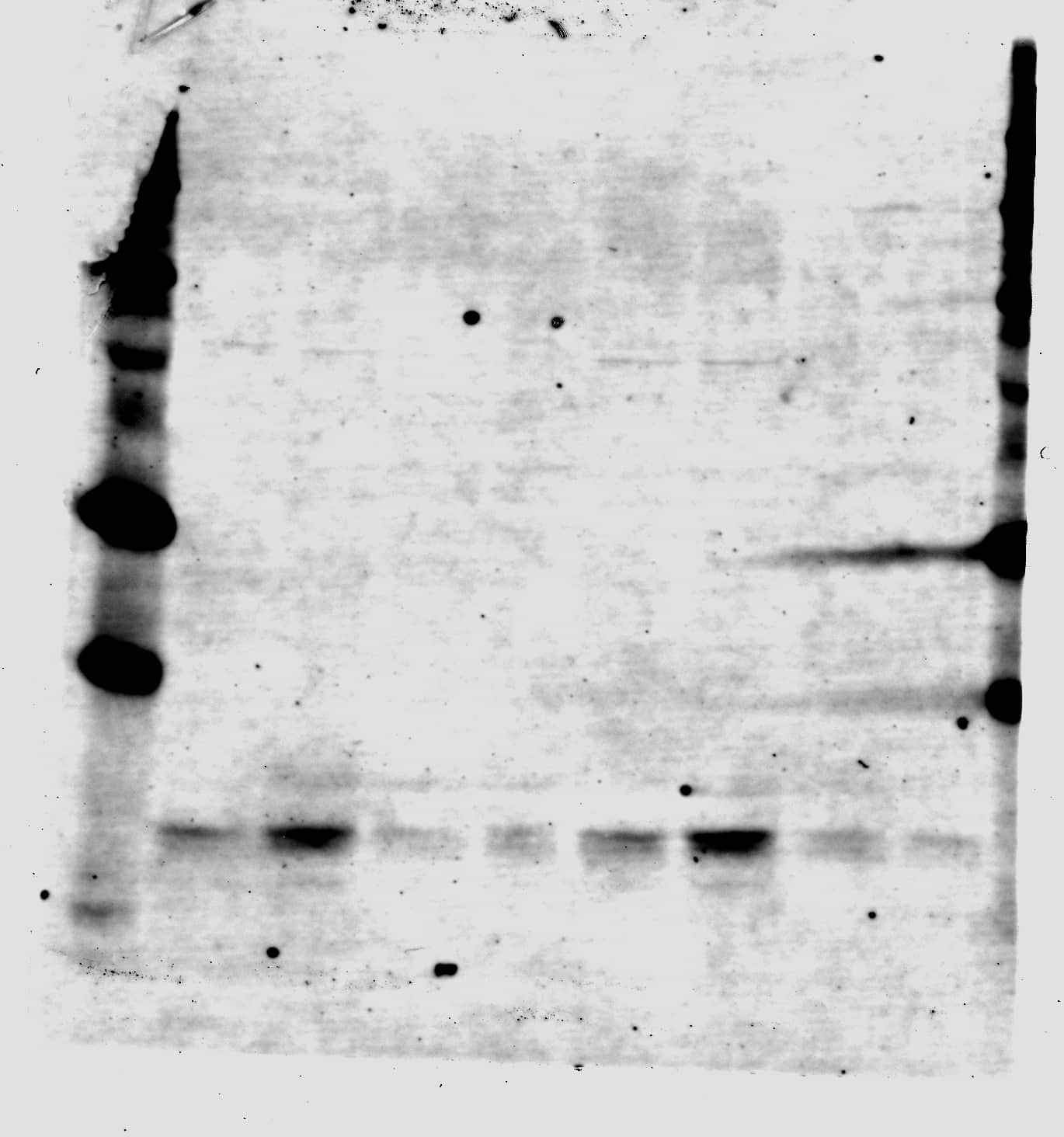
![Western Blot: mCherry Antibody (1C51) [NBP1-96752] - WB assay of the crude extract of HEK293 cells transfected with pFin-EF1-mCherry vector (lane +) and an equal amount of protein extract from untransfected HEK293 cells (lane -). NBP1-96752 binds a major band running at about 28 kDa (observed molecular weight) corresponding to intact full-length mCherry. The two other bands are clearly processed forms of mCherry as they are not present in non-transfected HEK293 cells.](http://images.novusbio.com/fullsize/mCherry-Antibody-1C51-Western-Blot-NBP1-96752-img0002.jpg "Western Blot: mCherry Antibody (1C51) [NBP1-96752] - WB assay of the crude extract of HEK293 cells transfected with pFin-EF1-mCherry vector (lane +) and an equal amount of protein extract from untransfected HEK293 cells (lane -). NBP1-96752 binds a major band running at about 28 kDa (observed molecular weight) corresponding to intact full-length mCherry. The two other bands are clearly processed forms of mCherry as they are not present in non-transfected HEK293 cells.")
![Immunohistochemistry-Frozen: mCherry Antibody (1C51) [NBP1-96752] - Mouse Bone Marrow Sections (Femur). Fixed-frozen and decalcified. tdTomato reporter transgenic mice. tdTomato in hematopoietic cells were detected by anti-mCherry antibody. Antibody is cross-reactive and works well for fixed-frozen bone marrow. Background is low. IHC-Fr image submitted by a verified customer review.](http://images.novusbio.com/fullsize/mCherry-Antibody-1C51-Immunohistochemistry-Frozen-NBP1-96752-img0004.jpg "Immunohistochemistry-Frozen: mCherry Antibody (1C51) [NBP1-96752] - Mouse Bone Marrow Sections (Femur). Fixed-frozen and decalcified. tdTomato reporter transgenic mice. tdTomato in hematopoietic cells were detected by anti-mCherry antibody. Antibody is cross-reactive and works well for fixed-frozen bone marrow. Background is low. IHC-Fr image submitted by a verified customer review.")
![Western Blot: mCherry Antibody (1C51) [NBP1-96752] - Mis18 alpha :Mis18 beta -hexamer mediates dimerization of M18BP1.(A) Analytical SEC results of M18BP11–140-MBP (cyan), M18BP11–228-MBP (red), Mis18 alpha :Mis18 beta :M18BP11–140-MBP (purple), Mis18 alpha :Mis18 beta :M18BP11–228-MBP (green). The elution volumes of thyroglobulin (670 kD), ferritin (440 kD), catalase (240 kD) & ovalbumin (44 kD) are shown as standards. Red lines indicate fractions collected for Tricine–SDS-PAGE analyses. Gels were stained with CBB. (B) Sedimentation velocity AUC results of the same samples used in the analytical SEC experiments (panel A). The best-fit size distributions are shown with the colors indicated in panel A. Data profiles used for curve-fitting analyses are shown in Figure 7—figure supplement 1. (C) Summary table of the results obtained from the AUC experiments of panel B. Sed. coef., sedimentation coefficient; MWobs., observed molecular weight; MWtheo., theoretical molecular weight. (D) Western blot results of co-immunoprecipitation experiments using GFP-Trap_A beads. HeLa CENP-A-SNAP + EGFP-M18BP11–140-P2A-T2A-mCherry-M18BP11–140, EGFP-M18BP11–140/T40D/S110E-P2A-T2A-mCherry-M18BP11–140/T40D/S110E, GST-EGFP-M18BP11–140-P2A-T2A-GST-mCherry-M18BP11–140, or GST-EGFP-M18BP11–140/T40D/S110E-P2A-T2A-GST-mCherry-M18BP11–140/T40D/S110E were analyzed.DOI://dx.doi.org/10.7554/eLife.23352.014Data profiles for AUC experiments.Best-fitting results of the sedimentation velocity AUC data of M18BP11–140-MBP, M18BP11–228-MBP, Mis18 alpha :Mis18 beta :M18BP11–140-MBP, and Mis18 alpha :Mis18 beta :M18BP11–228-MBP. Residuals represent the deviation of the continuous c(s) distribution model from the observed signals. The values of RMSD for data fitting are shown.DOI://dx.doi.org/10.7554/eLife.23352.015 Image collected & cropped by CiteAb from the following publication (//pubmed.ncbi.nlm.nih.gov/28059702), licensed under a CC-BY license. Not internally tested by Novus Biologicals.](http://images.novusbio.com/fullsize/nbp1-96752_mouse-monoclonal-mcherry-antibody-1c51-310202416113527.jpg "Western Blot: mCherry Antibody (1C51) [NBP1-96752] - Mis18 alpha :Mis18 beta -hexamer mediates dimerization of M18BP1.(A) Analytical SEC results of M18BP11–140-MBP (cyan), M18BP11–228-MBP (red), Mis18 alpha :Mis18 beta :M18BP11–140-MBP (purple), Mis18 alpha :Mis18 beta :M18BP11–228-MBP (green). The elution volumes of thyroglobulin (670 kD), ferritin (440 kD), catalase (240 kD) & ovalbumin (44 kD) are shown as standards. Red lines indicate fractions collected for Tricine–SDS-PAGE analyses. Gels were stained with CBB. (B) Sedimentation velocity AUC results of the same samples used in the analytical SEC experiments (panel A). The best-fit size distributions are shown with the colors indicated in panel A. Data profiles used for curve-fitting analyses are shown in Figure 7—figure supplement 1. (C) Summary table of the results obtained from the AUC experiments of panel B. Sed. coef., sedimentation coefficient; MWobs., observed molecular weight; MWtheo., theoretical molecular weight. (D) Western blot results of co-immunoprecipitation experiments using GFP-Trap_A beads. HeLa CENP-A-SNAP + EGFP-M18BP11–140-P2A-T2A-mCherry-M18BP11–140, EGFP-M18BP11–140/T40D/S110E-P2A-T2A-mCherry-M18BP11–140/T40D/S110E, GST-EGFP-M18BP11–140-P2A-T2A-GST-mCherry-M18BP11–140, or GST-EGFP-M18BP11–140/T40D/S110E-P2A-T2A-GST-mCherry-M18BP11–140/T40D/S110E were analyzed.DOI://dx.doi.org/10.7554/eLife.23352.014Data profiles for AUC experiments.Best-fitting results of the sedimentation velocity AUC data of M18BP11–140-MBP, M18BP11–228-MBP, Mis18 alpha :Mis18 beta :M18BP11–140-MBP, and Mis18 alpha :Mis18 beta :M18BP11–228-MBP. Residuals represent the deviation of the continuous c(s) distribution model from the observed signals. The values of RMSD for data fitting are shown.DOI://dx.doi.org/10.7554/eLife.23352.015 Image collected & cropped by CiteAb from the following publication (//pubmed.ncbi.nlm.nih.gov/28059702), licensed under a CC-BY license. Not internally tested by Novus Biologicals.")
![Western Blot: mCherry Antibody (1C51) [NBP1-96752] - The oligomerization state of overexpressed Cav1 varies as a function of its tag. COS-7 cells expressing the indicated constructs were lysed in digitonin & subjected to BN-PAGE followed by western blotting for Cav1 (red) & either GFP, mCherry or myc (green). A) Cells were either left untransfected (‘control’) or transfected with EGFP, Cav1-GFP or P132L-GFP. B) As in (A) except cells were transfected with the indicated mCherry constructs. C) As in (A) except cells were transfected with Cav1-myc or P132L-myc. Figures are representative of two independent experiments. Red arrows indicate the high molecular weight band positive for both tag antibodies & Cav1 antibodies (h1-97 or 2297). Black arrows indicate the high molecular weight band only positive for Cav1 antibodies (h1-97 or 2297). Green arrows indicate the low molecular weight bands only positive for FP tag antibodies. Image collected & cropped by CiteAb from the following publication (//pubmed.ncbi.nlm.nih.gov/25639341), licensed under a CC-BY license. Not internally tested by Novus Biologicals.](http://images.novusbio.com/fullsize/nbp1-96752_mouse-monoclonal-mcherry-antibody-1c51-3102024161220.jpg "Western Blot: mCherry Antibody (1C51) [NBP1-96752] - The oligomerization state of overexpressed Cav1 varies as a function of its tag. COS-7 cells expressing the indicated constructs were lysed in digitonin & subjected to BN-PAGE followed by western blotting for Cav1 (red) & either GFP, mCherry or myc (green). A) Cells were either left untransfected (‘control’) or transfected with EGFP, Cav1-GFP or P132L-GFP. B) As in (A) except cells were transfected with the indicated mCherry constructs. C) As in (A) except cells were transfected with Cav1-myc or P132L-myc. Figures are representative of two independent experiments. Red arrows indicate the high molecular weight band positive for both tag antibodies & Cav1 antibodies (h1-97 or 2297). Black arrows indicate the high molecular weight band only positive for Cav1 antibodies (h1-97 or 2297). Green arrows indicate the low molecular weight bands only positive for FP tag antibodies. Image collected & cropped by CiteAb from the following publication (//pubmed.ncbi.nlm.nih.gov/25639341), licensed under a CC-BY license. Not internally tested by Novus Biologicals.")

 followed by 30 min incubation with Goat anti Mouse HRP conjugated secondary antibodies (Catalog # HAF007) at 1:20 dilution + DAB chromogen (brown). The tissue was counterstained with Hematoxylin (blue). Control was done by omitting primary antibody.")

