HAF017). Specific bands were detected for VEGF-C at approximately 52 kDa, 34 kDa, and 13 kDa (as indicated). This experiment was conducted under reducing conditions and using
Immunoblot Buffer Group 1." class="big_lightbox">
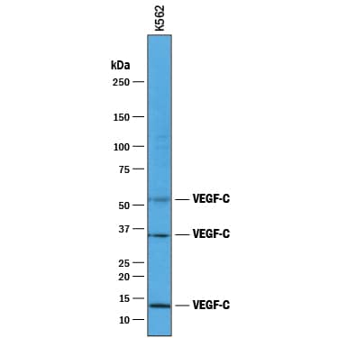
HAF017). Specific bands were detected for VEGF-C at approximately 52 kDa, 34 kDa, and 13 kDa (as indicated). This experiment was conducted under reducing conditions and using
Immunoblot Buffer Group 1." alt="Western blot shows lysates of K562 human chronic myelogenous leukemia cell line. PVDF membrane was probed with 1 µg/mL of Goat Anti-Human VEGF-C Antigen Affinity-purified Polyclonal Antibody (Catalog # AF752) followed by HRP-conjugated Anti-Goat IgG Secondary Antibody (
HAF017). Specific bands were detected for VEGF-C at approximately 52 kDa, 34 kDa, and 13 kDa (as indicated). This experiment was conducted under reducing conditions and using
Immunoblot Buffer Group 1." class="big_thumb" />
CTS008) and counterstained with hematoxylin (blue). Specific labeling was localized to epithelial cells in crypts of the colon mucusa (longitudinal section of crypts). Lower panel shows a lack of labeling if primary antibodies are omitted and tissue is stained only with secondary antibody followed by incubation with detection reagents. View our protocol for
Chromogenic IHC Staining of Paraffin-embedded Tissue Sections." class="big_lightbox">
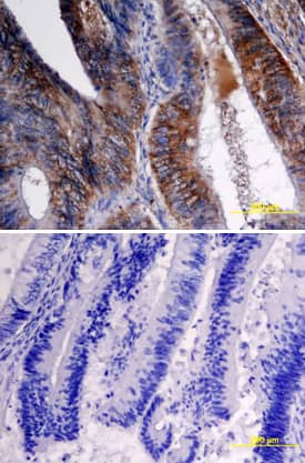
CTS008) and counterstained with hematoxylin (blue). Specific labeling was localized to epithelial cells in crypts of the colon mucusa (longitudinal section of crypts). Lower panel shows a lack of labeling if primary antibodies are omitted and tissue is stained only with secondary antibody followed by incubation with detection reagents. View our protocol for
Chromogenic IHC Staining of Paraffin-embedded Tissue Sections." class="big_thumb" />
MAB752) coated on a Clear Polystyrene Microplate (
DY990). Goat Anti-Human VEGF‑C Antigen Affinity-purified Polyclonal Antibody (Catalog # AF752) was biotinylated and incubated with the protein captured on the plate. Detection of the standard curve was achieved by incubating Streptavidin-HRP (
DY998) followed by Substrate Solution (
DY999) and stopping the enzymatic reaction with Stop Solution (
DY994)." class="big_lightbox">
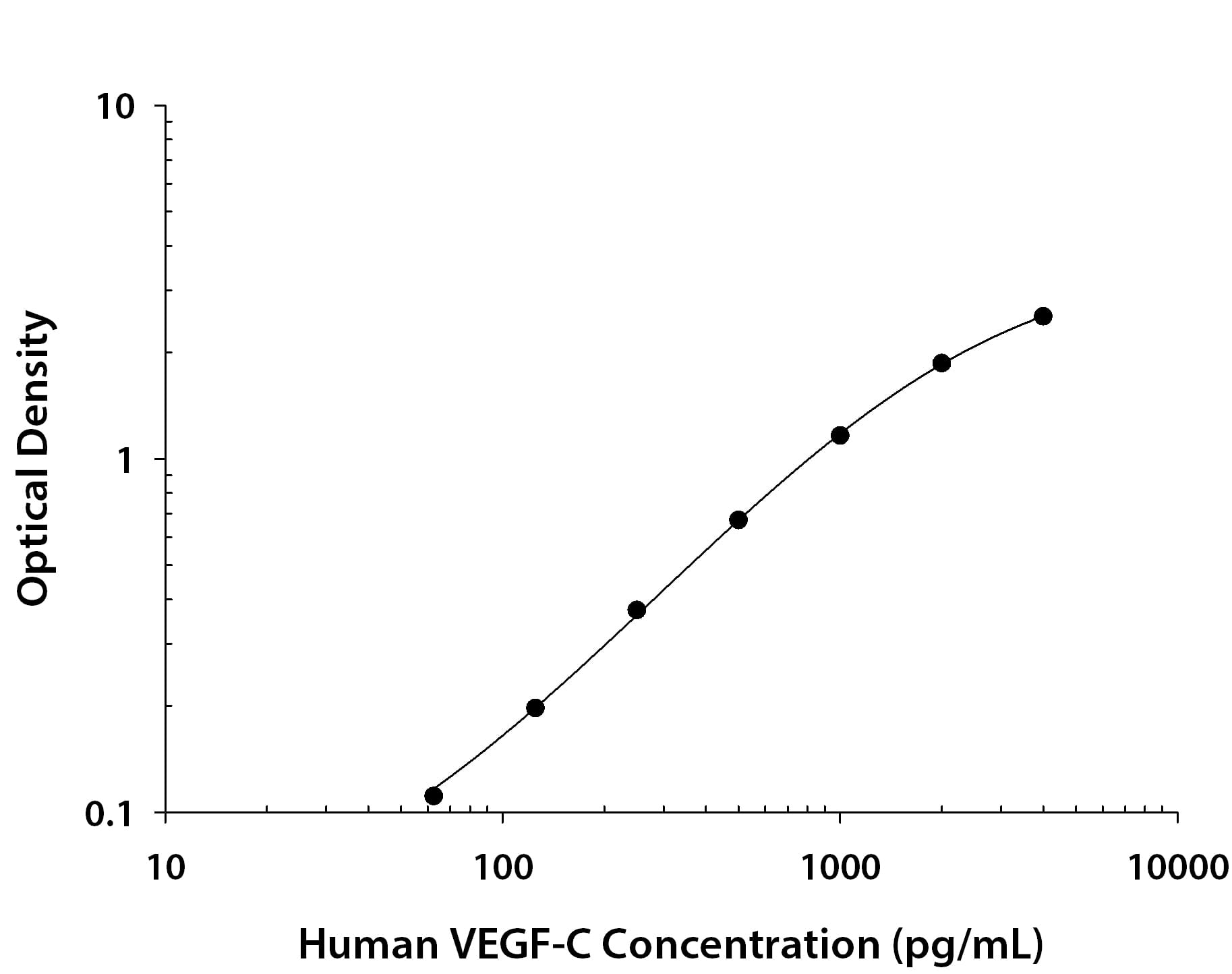
MAB752) coated on a Clear Polystyrene Microplate (
DY990). Goat Anti-Human VEGF‑C Antigen Affinity-purified Polyclonal Antibody (Catalog # AF752) was biotinylated and incubated with the protein captured on the plate. Detection of the standard curve was achieved by incubating Streptavidin-HRP (
DY998) followed by Substrate Solution (
DY999) and stopping the enzymatic reaction with Stop Solution (
DY994)." class="big_thumb" />
752-VC) was serially diluted and captured by Mouse Anti-Human VEGF‑C Monoclonal Antibody (Catalog #
MAB752) coated on a Clear Polystyrene Microplate (Catalog #
DY990). Goat Anti-Human VEGF‑C Antigen Affinity-purified Polyclonal Antibody (Catalog # AF752) was biotinylated and incubated with the protein captured on the plate. Detection of the standard curve was achieved by incubating Streptavidin-HRP (Catalog #
DY998)" class="big_lightbox">

752-VC) was serially diluted and captured by Mouse Anti-Human VEGF‑C Monoclonal Antibody (Catalog #
MAB752) coated on a Clear Polystyrene Microplate (Catalog #
DY990). Goat Anti-Human VEGF‑C Antigen Affinity-purified Polyclonal Antibody (Catalog # AF752) was biotinylated and incubated with the protein captured on the plate. Detection of the standard curve was achieved by incubating Streptavidin-HRP (Catalog #
DY998)" class="big_thumb" />
 DY990). Goat Anti-Human VEGF‑C Antigen Affinity-purified Polyclonal Antibody (AF752) was biotinylated and incubated with the protein captured on the plate. Detection of the standard curve was achieved by incubating Streptavidin-HRP (DY998) followed by Substrate Solution (DY999) and stopping the enzymatic reaction with Stop Solution (DY994)." alt="Recombinant Human VEGF‑C protein was serially diluted 2-fold and captured by Mouse Anti-Human VEGF‑C Monoclonal Antibody (Catalog # MAB752) coated on a Clear Polystyrene Microplate (DY990). Goat Anti-Human VEGF‑C Antigen Affinity-purified Polyclonal Antibody (AF752) was biotinylated and incubated with the protein captured on the plate. Detection of the standard curve was achieved by incubating Streptavidin-HRP (DY998) followed by Substrate Solution (DY999) and stopping the enzymatic reaction with Stop Solution (DY994)." class="big_thumb" />
 HAF017). Specific bands were detected for VEGF-C at approximately 52 kDa, 34 kDa, and 13 kDa (as indicated). This experiment was conducted under reducing conditions and using Immunoblot Buffer Group 1." alt="Western blot shows lysates of K562 human chronic myelogenous leukemia cell line. PVDF membrane was probed with 1 µg/mL of Goat Anti-Human VEGF-C Antigen Affinity-purified Polyclonal Antibody (Catalog # AF752) followed by HRP-conjugated Anti-Goat IgG Secondary Antibody (HAF017). Specific bands were detected for VEGF-C at approximately 52 kDa, 34 kDa, and 13 kDa (as indicated). This experiment was conducted under reducing conditions and using Immunoblot Buffer Group 1." class="big_thumb" />


![Immunocytochemistry/Immunofluorescence: VEGF-C Antibody [NB110-61022] - bEnd.3 mouse brain endothelioma cell line stained with Vegf-c antibody at 1:500. ICC/IF image submitted by a verified customer review.](http://images.novusbio.com/fullsize/VEGF-C-Antibody-Immunocytochemistry-Immunofluorescence-NB110-61022-img0002.jpg "Immunocytochemistry/Immunofluorescence: VEGF-C Antibody [NB110-61022] - bEnd.3 mouse brain endothelioma cell line stained with Vegf-c antibody at 1:500. ICC/IF image submitted by a verified customer review.")

![Immunohistochemistry: VEGF-C Antibody [NB110-61022] - VEGFC Antibody [NB110-61022] - 4% PFA fixed and paraffin embedded mouse Lung tissue section was subjected to IHC staining of mouse VEGF-C.](http://images.novusbio.com/fullsize/VEGF-C-Antibody-Immunohistochemistry-NB110-61022-img0001.jpg)
![Western Blot: VEGF-C Antibody [NB110-61022] - Kallikrein-related peptidase 3 (KLK3)/Prostate specific antigen (PSA) activates VEGF-C.(A, B) Cleavage of pro-VEGF-C by KLK3 (PSA). Pro-VEGF-C was incubated with or without KLK3, with & without the monoclonal antibody against KLK3 (5C7). Detection of VEGF-C in Western blots probed with antiserum 6 & 3/4, resulting in the detection of pro-VEGF-C (29/31 kDa) & activated, mature VEGF-C (21/23 kDa). The band marked by the asterisk likely represents the N-terminal propeptide (~15 kDa) which is detected by the antiserum 6. Note that for the image shown for antiserum 6, two different exposures of the same blot were merged (n = 3). (C, D) VEGF-C processed by KLK3 is biologically active in Ba/F3 cell assays, which translate activation of a hybrid VEGFR/EpoR receptor into cell survival (n = 2). Error bars indicate SD.10.7554/eLife.44478.004Figure 1—source data 1.Ba/F3 assay showing the activity of KLK3-cleaved VEGF-C.Ba/F3 assay showing the activity of KLK3-cleaved VEGF-C. Image collected & cropped by CiteAb from the following publication (//pubmed.ncbi.nlm.nih.gov/31099754), licensed under a CC-BY license. Not internally tested by Novus Biologicals.](http://images.novusbio.com/fullsize/nb110-61022_rabbit-polyclonal-vegf-c-antibody-31020241620375.jpg)

![Western Blot: VEGF-C Antibody (107/F10) [NBP1-18626] - Analysis of recombinant human VEGF-C derived from insect cells using a monoclonal antibody directed against recombinant human/rat VEGF-C.](http://images.novusbio.com/fullsize/VEGF-C-Antibody-107-F10-Western-Blot-NBP1-18626-img0001.jpg)
![Immunocytochemistry/Immunofluorescence: VEGF-C Antibody (107/F10) [NBP1-18626] - Human myofibroblasts stained with VEGF-C antibody. ICC/IF image submitted by a verified customer review.](http://images.novusbio.com/fullsize/VEGF-C-Antibody-107-F10-Immunocytochemistry-Immunofluorescence-NBP1-18626-img0003.jpg)
![Immunohistochemistry-Paraffin: VEGF-C Antibody (107/F10) [NBP1-18626] - Human gastric cancer tissue section stained with VEGF-C antibody. IHC-P image submitted by a verified customer review.](http://images.novusbio.com/fullsize/VEGF-C-Antibody-107-F10-Immunohistochemistry-Paraffin-NBP1-18626-img0002.jpg)
![Immunohistochemistry: VEGF-C Antibody (107/F10) - Azide and BSA Free [NBP1-18626] - Internal thoracic artery wall harvested from a patient diagnosed with double-vessel CAD showing immunohistochemical staining. Immunohistochemical staining of VEGF-C (a), CAV2 (b) & CAV3 (c) in an internal thoracic artery obtained from a 67-year-old patient who developed the early graft restenosis within 7 months after coronary artery bypass grafting. VEGF-C is expressed in smooth muscle cells of the tunica media (TM) & endothelial cells present in the tunica intima (TI). CAV2 is present exclusively in smooth muscle cells. Strong expression of CAV3 is observed in smooth muscle cells & in endothelial cells. d Negative control. TA tunica adventitia Image collected & cropped by CiteAb from the following publication (//link.springer.com/10.1007/s00380-018-1158-9), licensed under a CC-BY license. Not internally tested by Novus Biologicals.](http://images.novusbio.com/fullsize/nbp1-18626_mouse-monoclonal-vegf-c-antibody-107-f10-31020241539259.jpg)
![Immunohistochemistry: VEGF-C Antibody (107/F10) - Azide and BSA Free [NBP1-18626] - Immunohistochemical staining of a saphenous vein wall harvested from a patient diagnosed with triple-vessel CAD. Three serial histological sections of a graft wall obtained from a 71-year-old patient who developed early graft failure 11 months after coronary artery bypass grafting (the same patients as mentioned in Fig. 7). VEGF-C (a), VEGFR-3 (b), & CAV3 (c) shown to be expressed by immunohistochemical staining in smooth muscle cells of the tunica media (TM). In addition, VEGFR-3 is strongly present in endothelial cells of the tunica intima (TI). d Negative control. TA tunica adventitia Image collected & cropped by CiteAb from the following publication (//link.springer.com/10.1007/s00380-018-1158-9), licensed under a CC-BY license. Not internally tested by Novus Biologicals.](http://images.novusbio.com/fullsize/nbp1-18626_mouse-monoclonal-vegf-c-antibody-107-f10-310202415371967.jpg)
![Immunohistochemistry: VEGF-C Antibody (107/F10) - Azide and BSA Free [NBP1-18626] - Immunohistochemical staining of an internal thoracic artery & saphenous vein wall harvested from a patient with no evidence of luminal stenosis 12 months after CABG. Immunohistochemical staining of VEGF-C (a) & CAV2 (b) in an internal thoracic artery (ITA) graft & VEGFR-3 (c) in saphenous vein (SV) transplant harvested from a 65-year-old patient diagnosed with triple-vessel CAD. Note that lack of CAV2 expression within smooth muscle cells (SMC) in ITA wall is accompanied by VEGF-C positive expression in the same area. In SV tissue sample harvested from the same patient, no VEGRR-3 expression was found. d Negative control. TI tunica intima, TM tunica media, TA tunica adventitia Image collected & cropped by CiteAb from the following publication (//link.springer.com/10.1007/s00380-018-1158-9), licensed under a CC-BY license. Not internally tested by Novus Biologicals.](http://images.novusbio.com/fullsize/nbp1-18626_mouse-monoclonal-vegf-c-antibody-107-f10-31020241535674.jpg)
![Immunohistochemistry: VEGF-C Antibody (107/F10) - Azide and BSA Free [NBP1-18626] - Saphenous vein wall harvested from a patient diagnosed with double-vessel CAD showing immunohistochemical staining. VEGF-C (a) & VEGFR-3 (b) reactivity in a graft wall obtained from a 67-year-old patient who developed the early graft restenosis within 7 months after coronary artery bypass grafting (the same patient as mentioned in Fig. 6). VEGF-C is localized exclusively within individual smooth muscle cells of the tunica media (TM). Cells with positive VEGFR-3 expression are situated in endothelial cells of the tunica intima (TI). c Negative control. TA tunica adventitia Image collected & cropped by CiteAb from the following publication (//link.springer.com/10.1007/s00380-018-1158-9), licensed under a CC-BY license. Not internally tested by Novus Biologicals.](http://images.novusbio.com/fullsize/nbp1-18626_mouse-monoclonal-vegf-c-antibody-107-f10-310202415395931.jpg)
![Immunohistochemistry: VEGF-C Antibody (107/F10) - Azide and BSA Free [NBP1-18626] - Immunohistochemistry of an internal thoracic artery wall harvested from a patient diagnosed with triple-vessel CAD. Immunohistochemical staining of VEGF-C (a), CAV2 (b), & CAV3 (c) in an internal thoracic artery graft harvested from a 71-year-old patient who developed early graft failure 11 months after coronary artery bypass grafting. Intensive VEGF-C & CAV3 expression is observed in the smooth muscle cells of the tunica media (TM) & in the endothelial cells of the tunica intima (TI). The presence of immunopositive expression of CAV2 (c) is limited to smooth muscle cells of the tunica media (TM). Expression of all analyzed proteins is present in the small blood vessels localized in the tunica adventitia (TA, arrows). d Negative control Image collected & cropped by CiteAb from the following publication (//link.springer.com/10.1007/s00380-018-1158-9), licensed under a CC-BY license. Not internally tested by Novus Biologicals.](http://images.novusbio.com/fullsize/nbp1-18626_mouse-monoclonal-vegf-c-antibody-107-f10-310202416165622.jpg)
![Immunohistochemistry-Paraffin: VEGF-C Antibody [NBP2-20853] - Human hepatoma, using VEGFC antibody at 1:500 dilution. Antigen Retrieval: Trilogy™ (EDTA based, pH 8.0) buffer, 15min](http://images.novusbio.com/fullsize/VEGFC-Antibody-Immunohistochemistry-Paraffin-NBP2-20853-img0003.jpg "Immunohistochemistry-Paraffin: VEGF-C Antibody [NBP2-20853] - Human hepatoma, using VEGFC antibody at 1:500 dilution. Antigen Retrieval: Trilogy™ (EDTA based, pH 8.0) buffer, 15min")
![<b>Genetic Strategies Validation. </b>Western Blot: VEGF-C Antibody [NBP2-20853] - Sample (20 ug of whole cell lysate) A: TL1 whole cell lysate B: TL1 knock down VEGF-C whole cell lysate 12% SDS PAGE gel, diluted at 1:1000.](http://images.novusbio.com/fullsize/VEGFC-Antibody-Western-Blot-NBP2-20853-img0001.jpg)
![Western Blot: VEGF-C Antibody [NBP2-20853] - 50 ug mouse lung lysate/extract 12% SDS-PAGE VEGFC antibody dilution: 1:1000.](http://images.novusbio.com/fullsize/VEGFC-Antibody-Western-Blot-NBP2-20853-img0002.jpg)
![Western Blot: VEGF-C Antibody [NBP2-20853] - U87-MG whole cell extract and conditioned medium (30 ug) were separated by 10% SDS-PAGE, and the membrane was blotted with VEGF-C antibody (NBP2-20853) diluted at 1:500. The HRP-conjugated anti-rabbit IgG antibody was used to detect the primary antibody.](http://images.novusbio.com/fullsize/nbp2-20853_rabbit-polyclonal-vegf-c-antibody-910202420104436.jpg)


![Western Blot: VEGF-C Antibody (OTI4A1) [NBP2-45404] - Analysis of HEK293T cells were transfected with the pCMV6-ENTRY control (Left lane) or pCMV6-ENTRY VEGF-C.](http://images.novusbio.com/fullsize/VEGF-C-Antibody-OTI4A1-Western-Blot-NBP2-45404-img0001.jpg "Western Blot: VEGF-C Antibody (OTI4A1) [NBP2-45404] - Analysis of HEK293T cells were transfected with the pCMV6-ENTRY control (Left lane) or pCMV6-ENTRY VEGF-C.")
![Immunohistochemistry-Paraffin: VEGF-C Antibody (OTI4A1) [NBP2-45404] - Staining of paraffin-embedded Human prostate tissue within the normal limits using anti-VEGFC mouse monoclonal antibody. (Heat-induced epitope retrieval by 1mM EDTA in 10mM Tris buffer (pH8.5) at 120C for 3min) (1:150)](http://images.novusbio.com/fullsize/VEGF-C-Antibody-OTI4A1-Immunohistochemistry-Paraffin-NBP2-45404-img0008.jpg)
![Immunohistochemistry-Paraffin: VEGF-C Antibody (OTI4A1) [NBP2-45404] - Staining of paraffin-embedded Carcinoma of Human liver tissue using anti-VEGFC mouse monoclonal antibody. (Heat-induced epitope retrieval by 1mM EDTA in 10mM Tris buffer (pH8.5) at 120C for 3min) (1:150)](http://images.novusbio.com/fullsize/VEGF-C-Antibody-OTI4A1-Immunohistochemistry-Paraffin-NBP2-45404-img0002.jpg)
![Immunohistochemistry-Paraffin: VEGF-C Antibody (OTI4A1) [NBP2-45404] - Staining of paraffin-embedded Carcinoma of Human pancreas tissue using anti-VEGFC mouse monoclonal antibody. (Heat-induced epitope retrieval by 1mM EDTA in 10mM Tris buffer (pH8.5) at 120C for 3min) (1:150)](http://images.novusbio.com/fullsize/VEGF-C-Antibody-OTI4A1-Immunohistochemistry-Paraffin-NBP2-45404-img0003.jpg)
![Immunohistochemistry-Paraffin: VEGF-C Antibody (OTI4A1) [NBP2-45404] - Staining of paraffin-embedded Carcinoma of Human prostate tissue using anti-VEGFC mouse monoclonal antibody. (Heat-induced epitope retrieval by 1mM EDTA in 10mM Tris buffer (pH8.5) at 120C for 3min) (1:150)](http://images.novusbio.com/fullsize/VEGF-C-Antibody-OTI4A1-Immunohistochemistry-Paraffin-NBP2-45404-img0004.jpg)
![Immunohistochemistry-Paraffin: VEGF-C Antibody (OTI4A1) [NBP2-45404] - Staining of paraffin-embedded Human endometrium tissue within the normal limits using anti-VEGFC mouse monoclonal antibody. (Heat-induced epitope retrieval by 1mM EDTA in 10mM Tris buffer (pH8.5) at 120C for 3min) (1:150)](http://images.novusbio.com/fullsize/VEGF-C-Antibody-OTI4A1-Immunohistochemistry-Paraffin-NBP2-45404-img0005.jpg)
![Immunohistochemistry-Paraffin: VEGF-C Antibody (OTI4A1) [NBP2-45404] - Staining of paraffin-embedded Human liver tissue within the normal limits using anti-VEGFC mouse monoclonal antibody. (Heat-induced epitope retrieval by 1mM EDTA in 10mM Tris buffer (pH8.5) at 120C for 3min) (1:150)](http://images.novusbio.com/fullsize/VEGF-C-Antibody-OTI4A1-Immunohistochemistry-Paraffin-NBP2-45404-img0006.jpg)
![Immunohistochemistry-Paraffin: VEGF-C Antibody (OTI4A1) [NBP2-45404] - Staining of paraffin-embedded Human pancreas tissue within the normal limits using anti-VEGFC mouse monoclonal antibody. (Heat-induced epitope retrieval by 1mM EDTA in 10mM Tris buffer (pH8.5) at 120C for 3min) (1:150)](http://images.novusbio.com/fullsize/VEGF-C-Antibody-OTI4A1-Immunohistochemistry-Paraffin-NBP2-45404-img0007.jpg)
![Immunohistochemistry-Paraffin: VEGF-C Antibody (OTI4A1) [NBP2-45404] - Staining of paraffin-embedded Human tonsil within the normal limits using anti-VEGFC mouse monoclonal antibody. (Heat-induced epitope retrieval by 1mM EDTA in 10mM Tris buffer (pH8.5) at 120C for 3min) (1:150)](http://images.novusbio.com/fullsize/VEGF-C-Antibody-OTI4A1-Immunohistochemistry-Paraffin-NBP2-45404-img0009.jpg)
 752-VC) was serially diluted and captured by Mouse Anti-Human VEGF‑C Monoclonal Antibody (Catalog # MAB752) coated on a Clear Polystyrene Microplate (Catalog #
 ![Western Blot: VEGF-C Antibody (9/G10) - Azide and BSA Free [NBP2-36287] - Western Blot analysis of recombinant human VEGF-C derived from insect cells using a monoclonal antibody directed against recombinant human/rat VEGF-C. The antibody does not recognize the unreduced form. The bands correspond to different glycosylated forms.](http://images.novusbio.com/fullsize/VEGF-C-Antibody-9-G10-Azide-and-BSA-Free-Western-Blot-NBP2-36287-img0008.jpg "Western Blot: VEGF-C Antibody (9/G10) - Azide and BSA Free [NBP2-36287] - Western Blot analysis of recombinant human VEGF-C derived from insect cells using a monoclonal antibody directed against recombinant human/rat VEGF-C. The antibody does not recognize the unreduced form. The bands correspond to different glycosylated forms.")
![Western Blot: VEGF-C Antibody (9/G10) [NBP2-36287] - Analysis of recombinant rat VEGF-C derived from insect cells using a monoclonal antibody directed against recombinant human/rat VEGF-C. The antibody does not recognize the unreduced form. The bands correspond to different glycosylated forms.](http://images.novusbio.com/fullsize/VEGF-C-Antibody-9-G10-Western-Blot-NBP2-36287-img0006.jpg)
![Sandwich ELISA Capture: VEGF-C Antibody (9/G10) [NBP2-36287] - VEGF-C Sandwich-ELISA using recombinant human VEGF-C as standard. Mouse anti-human VEGF-C #9/G10 was used as capture antibody, Biotinylated mouse anti-human VEGF was used for detection.](http://images.novusbio.com/fullsize/VEGF-C-Antibody-9-G10-Sandwich-ELISA-Capture-NBP2-36287-img0002.jpg)
![ELISA: VEGF-C Antibody (9/G10) - Azide and BSA Free [NBP2-36287] - VEGF-C Sandwich-ELISA using recombinant human VEGF-C as standard. Mouse anti-human VEGF-C #9/G10 was used as capture antibody, Biotinylated mouse anti-human VEGF-C was used for detection.](http://images.novusbio.com/fullsize/VEGF-C-Antibody-9-G10-Azide-and-BSA-Free-ELISA-NBP2-36287-img0007.jpg)
![Western Blot: VEGF-C Antibody (HL2289) - Azide and BSA Free [NBP3-25735] - U87-MG whole cell extract and conditioned medium (30 ug) were separated by 10% SDS-PAGE, and the membrane was blotted with VEGFC antibody [HL2289] (NBP3-25735) diluted at 1:1000. The HRP-conjugated anti-rabbit IgG antibody was used to detect the primary antibody.](http://images.novusbio.com/fullsize/nbp3-25735_rabbit-vegf-c-mab-hl2289-azide-and-bsa-free-2120249403891.jpg "Western Blot: VEGF-C Antibody (HL2289) - Azide and BSA Free [NBP3-25735] - U87-MG whole cell extract and conditioned medium (30 ug) were separated by 10% SDS-PAGE, and the membrane was blotted with VEGFC antibody [HL2289] (NBP3-25735) diluted at 1:1000. The HRP-conjugated anti-rabbit IgG antibody was used to detect the primary antibody.")
![Immunocytochemistry/Immunofluorescence: VEGF-C Antibody (HL2289) - Azide and BSA Free [NBP3-25735] - VEGFC antibody [HL2289] detects VEGFC protein at cytoplasmic vesicles by immunofluorescent analysis. Sample: U87-MG cells were fixed in 4% paraformaldehyde at RT for 15 min. Green: VEGFC stained by VEGFC antibody [HL2289] (NBP3-25735) diluted at 1:500. Red: alpha Tubulin, a cytoskeleton marker, stained by alpha Tubulin antibody [GT114] (NBP2-43837) diluted at 1:1000. Blue: Fluoroshield with DAPI .](http://images.novusbio.com/fullsize/nbp3-25735_rabbit-vegf-c-mab-hl2289-azide-and-bsa-free-21202494147.jpg)

![Sandwich ELISA: VEGF-C Antibody (107/A11) [NBP2-36288] - Recombinant human VEGF-C as standard. Mouse anti-human VEGF-C #9/G10 was used as capture antibody, Biotinylated mouse anti-human VEGF-C #107/A11 was used for detection.](http://images.novusbio.com/fullsize/VEGF-C-Antibody-107-A11-Sandwich-ELISA-NBP2-36288-img0001.jpg "Sandwich ELISA: VEGF-C Antibody (107/A11) [NBP2-36288] - Recombinant human VEGF-C as standard. Mouse anti-human VEGF-C #9/G10 was used as capture antibody, Biotinylated mouse anti-human VEGF-C #107/A11 was used for detection.")
![Immunohistochemistry-Frozen: VEGF-C Antibody (9/E7) [NBP1-18627] - Staining of VEGF-C on cryo sections (acetone-fixed) of rat mammary tumors.](http://images.novusbio.com/fullsize/VEGF-C-Antibody-9-E7-Immunohistochemistry-Frozen-NBP1-18627-img0006.jpg "Immunohistochemistry-Frozen: VEGF-C Antibody (9/E7) [NBP1-18627] - Staining of VEGF-C on cryo sections (acetone-fixed) of rat mammary tumors.")
 CTS008) and counterstained with hematoxylin (blue). Specific labeling was localized to stromal cells surrounding crypts in the colon mucosa (cross section across crypts). View our protocol for
  CTS008) and counterstained with hematoxylin (blue). Specific labeling was localized to epithelial cells in crypts of the colon mucusa (longitudinal section of crypts). Lower panel shows a lack of labeling if primary antibodies are omitted and tissue is stained only with secondary antibody followed by incubation with detection reagents. View our protocol for
  MAB752) coated on a Clear Polystyrene Microplate (

 752-VC) was serially diluted and captured by Mouse Anti-Human VEGF‑C Monoclonal Antibody (Catalog #


