1535-LM) reduces the Encephalomyocarditis Virus (EMCV)-induced cytopathy in the L-929 mouse fibroblast cell line in a dose-dependent manner (orange line), as measured by Resazurin (
AR002). Inhibition of EMCV activity elicited by Recombinant Mouse Limitin (30 pg/mL) is neutralized (green line) by increasing concentrations of Goat Anti-Mouse Limitin Antigen Affinity-purified Polyclonal Antibody (Catalog # AF597). The ND
50 is typically 1.5-7.5 ng/mL." class="big_lightbox">
1535-LM) reduces the Encephalomyocarditis Virus (EMCV)-induced cytopathy in the L-929 mouse fibroblast cell line in a dose-dependent manner (orange line), as measured by Resazurin (
AR002). Inhibition of EMCV activity elicited by Recombinant Mouse Limitin (30 pg/mL) is neutralized (green line) by increasing concentrations of Goat Anti-Mouse Limitin Antigen Affinity-purified Polyclonal Antibody (Catalog # AF597). The ND
50 is typically 1.5-7.5 ng/mL." alt="Recombinant Mouse Limitin (
1535-LM) reduces the Encephalomyocarditis Virus (EMCV)-induced cytopathy in the L-929 mouse fibroblast cell line in a dose-dependent manner (orange line), as measured by Resazurin (
AR002). Inhibition of EMCV activity elicited by Recombinant Mouse Limitin (30 pg/mL) is neutralized (green line) by increasing concentrations of Goat Anti-Mouse Limitin Antigen Affinity-purified Polyclonal Antibody (Catalog # AF597). The ND
50 is typically 1.5-7.5 ng/mL." class="big_thumb" />
VC004) or the HRP-conjugated Anti-Goat IgG Secondary Antibody (Catalog #
HAF109). Before incubation with the primary antibody, tissue was subjected to heat-induced epitope retrieval using VisUCyte Antigen Retrieval Reagent-Basic (Catalog #
VCTS021). Tissue was stained using DAB (brown) and counterstained with hematoxylin (blue). Specific staining was localized to the membrane of lymphocytes. View our protocol for
Chromogenic IHC Staining of Paraffin-embedded Tissue Sections." class="big_lightbox">
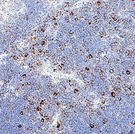
VC004) or the HRP-conjugated Anti-Goat IgG Secondary Antibody (Catalog #
HAF109). Before incubation with the primary antibody, tissue was subjected to heat-induced epitope retrieval using VisUCyte Antigen Retrieval Reagent-Basic (Catalog #
VCTS021). Tissue was stained using DAB (brown) and counterstained with hematoxylin (blue). Specific staining was localized to the membrane of lymphocytes. View our protocol for
Chromogenic IHC Staining of Paraffin-embedded Tissue Sections." class="big_thumb" />
1535-LM) was serially diluted and captured by Rat Anti-Mouse Limitin/IFN‑ zeta Monoclonal Antibody (Catalog #
MAB5971) coated on a Clear Polystyrene Microplate (Catalog #
DY990). Goat Anti-Mouse Limitin/IFN‑ zeta Antigen Affinity-purified Polyclonal Antibody (Catalog # AF597) was biotinylated and incubated with the protein captured on the plate. Detection of the standard curve was achieved by incubating Streptavidin-HRP (Catalog #
DY998)" class="big_lightbox">

1535-LM) was serially diluted and captured by Rat Anti-Mouse Limitin/IFN‑ zeta Monoclonal Antibody (Catalog #
MAB5971) coated on a Clear Polystyrene Microplate (Catalog #
DY990). Goat Anti-Mouse Limitin/IFN‑ zeta Antigen Affinity-purified Polyclonal Antibody (Catalog # AF597) was biotinylated and incubated with the protein captured on the plate. Detection of the standard curve was achieved by incubating Streptavidin-HRP (Catalog #
DY998)" class="big_thumb" />
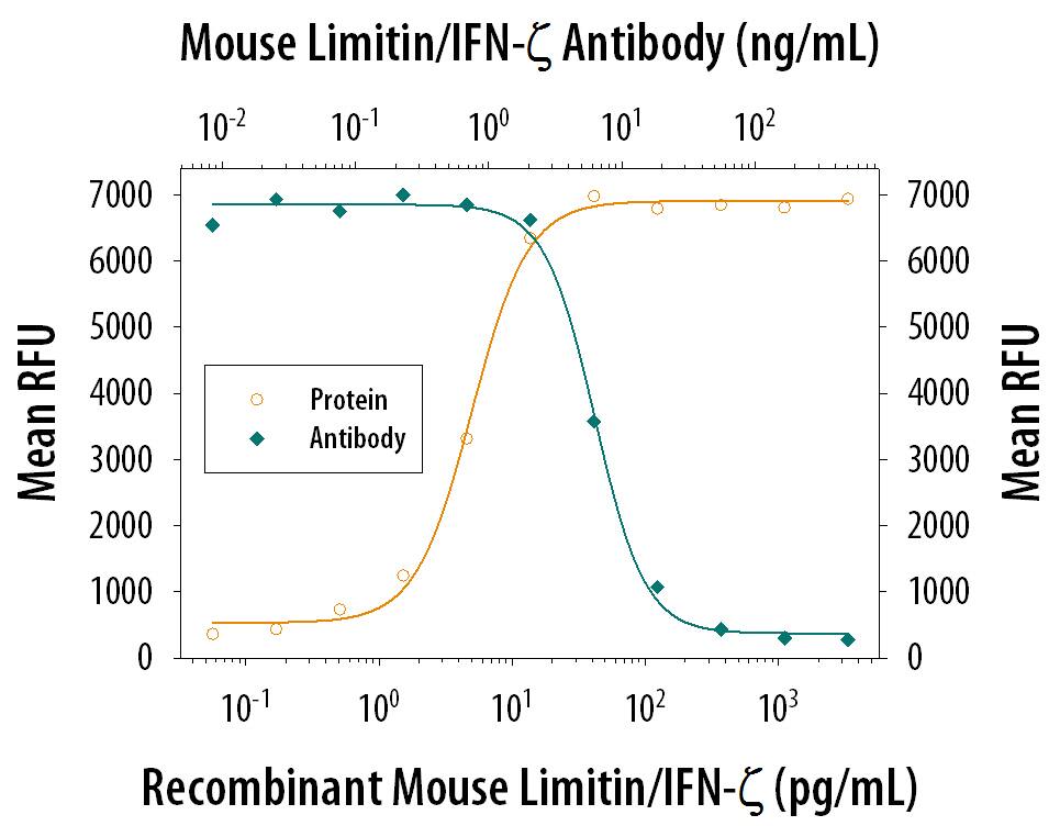
 1535-LM) reduces the Encephalomyocarditis Virus (EMCV)-induced cytopathy in the L-929 mouse fibroblast cell line in a dose-dependent manner (orange line), as measured by Resazurin (AR002). Inhibition of EMCV activity elicited by Recombinant Mouse Limitin (30 pg/mL) is neutralized (green line) by increasing concentrations of Goat Anti-Mouse Limitin Antigen Affinity-purified Polyclonal Antibody (Catalog # AF597). The ND50 is typically 1.5-7.5 ng/mL." alt="Recombinant Mouse Limitin (1535-LM) reduces the Encephalomyocarditis Virus (EMCV)-induced cytopathy in the L-929 mouse fibroblast cell line in a dose-dependent manner (orange line), as measured by Resazurin (AR002). Inhibition of EMCV activity elicited by Recombinant Mouse Limitin (30 pg/mL) is neutralized (green line) by increasing concentrations of Goat Anti-Mouse Limitin Antigen Affinity-purified Polyclonal Antibody (Catalog # AF597). The ND50 is typically 1.5-7.5 ng/mL." class="big_thumb" />
 1535-LM) was serially diluted and captured by Rat Anti-Mouse Limitin/IFN‑ zeta Monoclonal Antibody (Catalog # MAB5971) coated on a Clear Polystyrene Microplate (Catalog # DY990). Goat Anti-Mouse Limitin/IFN‑ zeta Antigen Affinity-purified Polyclonal Antibody (Catalog # AF597) was biotinylated and incubated with the protein captured on the plate. Detection of the standard curve was achieved by incubating Streptavidin-HRP (Catalog # DY998)" alt="Recombinant Mouse Limitin/IFN‑ zeta (Catalog # 1535-LM) was serially diluted and captured by Rat Anti-Mouse Limitin/IFN‑ zeta Monoclonal Antibody (Catalog # MAB5971) coated on a Clear Polystyrene Microplate (Catalog # DY990). Goat Anti-Mouse Limitin/IFN‑ zeta Antigen Affinity-purified Polyclonal Antibody (Catalog # AF597) was biotinylated and incubated with the protein captured on the plate. Detection of the standard curve was achieved by incubating Streptavidin-HRP (Catalog # DY998)" class="big_thumb" />


 VC004) or the HRP-conjugated Anti-Goat IgG Secondary Antibody (Catalog #
  1535-LM) was serially diluted and captured by Rat Anti-Mouse Limitin/IFN‑ zeta Monoclonal Antibody (Catalog #
 ![Immunohistochemistry-Paraffin: Limitin/IFN-zeta Antibody (RM0123-7D19) [NBP2-12426] - The Kidney tissue samples from the Folic Acid induced kidney injury model were fixed using 4% PFA at 4C for overnight and embeded in paraffin. 4um section was sublected to IHC with NBP2-12426](http://images.novusbio.com/fullsize/Limitin-IFN-zeta-Antibody-RM0123-7D19-Immunohistochemistry-Paraffin-NBP2-12426-img0001.jpg "Immunohistochemistry-Paraffin: Limitin/IFN-zeta Antibody (RM0123-7D19) [NBP2-12426] - The Kidney tissue samples from the Folic Acid induced kidney injury model were fixed using 4% PFA at 4C for overnight and embeded in paraffin. 4um section was sublected to IHC with NBP2-12426")


