VC001). Before incubation with the primary antibody, tissue was subjected to heat-induced epitope retrieval using Antigen Retrieval Reagent-Basic (
CTS013). Tissue was stained using DAB (brown) and counterstained with hematoxylin (blue). Specific staining was localized to cytoplasm. Staining was performed using our protocol for IHC Staining with VisUCyte HRP Polymer Detection Reagents." class="big_lightbox">
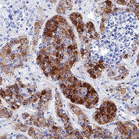
VC001). Before incubation with the primary antibody, tissue was subjected to heat-induced epitope retrieval using Antigen Retrieval Reagent-Basic (
CTS013). Tissue was stained using DAB (brown) and counterstained with hematoxylin (blue). Specific staining was localized to cytoplasm. Staining was performed using our protocol for IHC Staining with VisUCyte HRP Polymer Detection Reagents." alt="Neuronal Pentraxin 2 was detected in immersion fixed paraffin-embedded sections of human pituitary (anterior lobe) using Mouse Anti-Human Neuronal Pentraxin 2 Monoclonal Antibody (Catalog # MAB78162) at 0.5 µg/mL for 1 hour at room temperature followed by incubation with the Anti-Mouse IgG VisUCyte™ HRP Polymer Antibody (
VC001). Before incubation with the primary antibody, tissue was subjected to heat-induced epitope retrieval using Antigen Retrieval Reagent-Basic (
CTS013). Tissue was stained using DAB (brown) and counterstained with hematoxylin (blue). Specific staining was localized to cytoplasm. Staining was performed using our protocol for IHC Staining with VisUCyte HRP Polymer Detection Reagents." class="big_thumb" />
VC001). Before incubation with the primary antibody, tissue was subjected to heat-induced epitope retrieval using Antigen Retrieval Reagent-Basic (Catalog #
CTS013). Tissue was stained using DAB (brown) and counterstained with hematoxylin (blue). Specific staining was localized to cytoplasm. View our protocol for
IHC Staining with VisUCyte HRP Polymer Detection Reagents." class="big_lightbox">
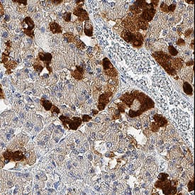
VC001). Before incubation with the primary antibody, tissue was subjected to heat-induced epitope retrieval using Antigen Retrieval Reagent-Basic (Catalog #
CTS013). Tissue was stained using DAB (brown) and counterstained with hematoxylin (blue). Specific staining was localized to cytoplasm. View our protocol for
IHC Staining with VisUCyte HRP Polymer Detection Reagents." class="big_thumb" />
VC001). Before incubation with the primary antibody, tissue was subjected to heat-induced epitope retrieval using Antigen Retrieval Reagent-Basic (
CTS013). Tissue was stained using DAB (brown) and counterstained with hematoxylin (blue). Specific staining was localized to islet cells. Staining was performed using our protocol for IHC Staining with VisUCyte HRP Polymer Detection Reagents." class="big_lightbox">
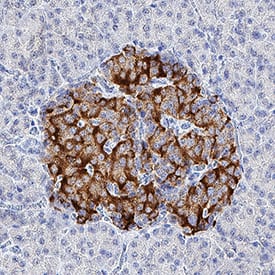
VC001). Before incubation with the primary antibody, tissue was subjected to heat-induced epitope retrieval using Antigen Retrieval Reagent-Basic (
CTS013). Tissue was stained using DAB (brown) and counterstained with hematoxylin (blue). Specific staining was localized to islet cells. Staining was performed using our protocol for IHC Staining with VisUCyte HRP Polymer Detection Reagents." class="big_thumb" />
MAB78161) coated on a Clear Polystyrene Microplate (Catalog #
DY990). Mouse Anti-Human Neuronal Pentraxin 2 Monoclonal Antibody (Catalog # MAB78162) was biotinylated and incubated with the protein captured on the plate. Detection of the standard curve was achieved by incubating Streptavidin-HRP (Catalog #
DY998) followed by Substrate Solution (Catalog #
DY999) and stopping the enzymatic reaction with Stop Solution (Catalog #
DY994)." class="big_lightbox">
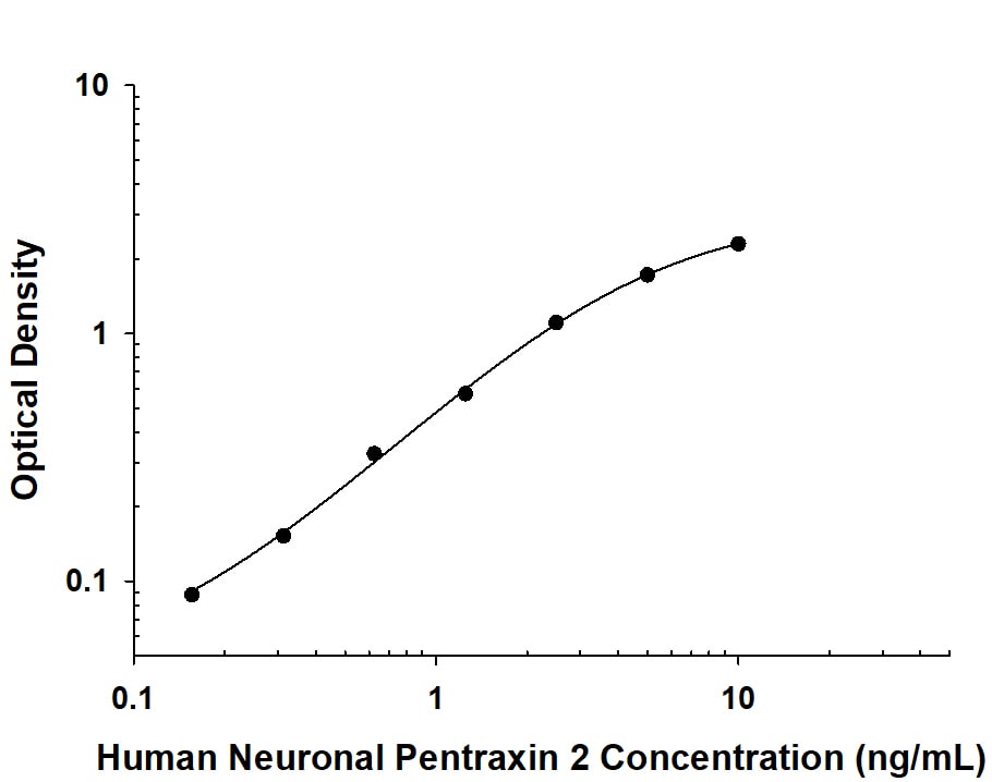
MAB78161) coated on a Clear Polystyrene Microplate (Catalog #
DY990). Mouse Anti-Human Neuronal Pentraxin 2 Monoclonal Antibody (Catalog # MAB78162) was biotinylated and incubated with the protein captured on the plate. Detection of the standard curve was achieved by incubating Streptavidin-HRP (Catalog #
DY998) followed by Substrate Solution (Catalog #
DY999) and stopping the enzymatic reaction with Stop Solution (Catalog #
DY994)." class="big_thumb" />
 VC001). Before incubation with the primary antibody, tissue was subjected to heat-induced epitope retrieval using Antigen Retrieval Reagent-Basic (CTS013). Tissue was stained using DAB (brown) and counterstained with hematoxylin (blue). Specific staining was localized to cytoplasm. Staining was performed using our protocol for IHC Staining with VisUCyte HRP Polymer Detection Reagents." alt="Neuronal Pentraxin 2 was detected in immersion fixed paraffin-embedded sections of human pituitary (anterior lobe) using Mouse Anti-Human Neuronal Pentraxin 2 Monoclonal Antibody (Catalog # MAB78162) at 0.5 µg/mL for 1 hour at room temperature followed by incubation with the Anti-Mouse IgG VisUCyte™ HRP Polymer Antibody (VC001). Before incubation with the primary antibody, tissue was subjected to heat-induced epitope retrieval using Antigen Retrieval Reagent-Basic (CTS013). Tissue was stained using DAB (brown) and counterstained with hematoxylin (blue). Specific staining was localized to cytoplasm. Staining was performed using our protocol for IHC Staining with VisUCyte HRP Polymer Detection Reagents." class="big_thumb" />
 NL007) and counterstained with DAPI (blue). Specific staining was localized to cytoplasm. View our protocol for Fluorescent ICC Staining of Cells on Coverslips." alt="Neuronal Pentraxin 2 was detected in immersion fixed SH-SY5Y human neuroblastoma cell line (left panel; positive staining) and U-251 MG human glioblastoma cell line (right panel; negative staining) using Mouse Anti-Human Neuronal Pentraxin 2 Monoclonal Antibody (Catalog # MAB7816) at 8 µg/mL for 3 hours at room temperature. Cells were stained using the NorthernLights™ 557-conjugated Anti-Mouse IgG Secondary Antibody (red; Catalog # NL007) and counterstained with DAPI (blue). Specific staining was localized to cytoplasm. View our protocol for Fluorescent ICC Staining of Cells on Coverslips." class="big_thumb" />
 VC001). Before incubation with the primary antibody, tissue was subjected to heat-induced epitope retrieval using Antigen Retrieval Reagent-Basic (CTS013). Tissue was stained using DAB (brown) and counterstained with hematoxylin (blue). Specific staining was localized to cytoplasm. View our protocol for IHC Staining with VisUCyte HRP Polymer Detection Reagents." alt="Neuronal Pentraxin 2 was detected in immersion fixed paraffin-embedded sections of human pituitary tissue using Mouse Anti-Human Neuronal Pentraxin 2 Monoclonal Antibody (Catalog # MAB78161) at 5 µg/mL for 1 hour at room temperature followed by incubation with the Anti-Mouse IgG VisUCyte™ HRP Polymer Antibody (VC001). Before incubation with the primary antibody, tissue was subjected to heat-induced epitope retrieval using Antigen Retrieval Reagent-Basic (CTS013). Tissue was stained using DAB (brown) and counterstained with hematoxylin (blue). Specific staining was localized to cytoplasm. View our protocol for IHC Staining with VisUCyte HRP Polymer Detection Reagents." class="big_thumb" />


![Western Blot: Neuronal Pentraxin 2 Antibody [NBP1-76899] - Independent Antibody Validation (IAV) via Protein Expression Profile in Cell Lines. Loading: 15 ug of lysates per lane. Antibodies: Neuronal Pentraxin 2 NBP1-76899 (2 ug/mL), Neuronal Pentraxin 2 (4 ug/mL), and beta-actin (1 ug/mL), 1h incubation at RT in 5% NFDM/TBST. Secondary: Goat anti-rabbit IgG HRP conjugate at 1:10000 dilution.](http://images.novusbio.com/fullsize/Neuronal-Pentraxin-2-Antibody-Western-Blot-NBP1-76899-img0011.jpg "Western Blot: Neuronal Pentraxin 2 Antibody [NBP1-76899] - Independent Antibody Validation (IAV) via Protein Expression Profile in Cell Lines. Loading: 15 ug of lysates per lane. Antibodies: Neuronal Pentraxin 2 NBP1-76899 (2 ug/mL), Neuronal Pentraxin 2 (4 ug/mL), and beta-actin (1 ug/mL), 1h incubation at RT in 5% NFDM/TBST. Secondary: Goat anti-rabbit IgG HRP conjugate at 1:10000 dilution.")
![Western Blot: Neuronal Pentraxin 2 Antibody [NBP1-76899] - Mouse brain tissue lysate with NPTX2 antibody at (A) 0.5 and (B) 1 ug/mL.](http://images.novusbio.com/fullsize/Neuronal-Pentraxin-2-Antibody-Western-Blot-NBP1-76899-img0001.jpg)
![Western Blot: Neuronal Pentraxin 2 Antibody [NBP1-76899] - Mouse brain tissue lysate with NPTX2 antibody at 1 ug/ml in (A) the absence and (B) the presence of blocking peptide.](http://images.novusbio.com/fullsize/Neuronal-Pentraxin-2-Antibody-Western-Blot-NBP1-76899-img0006.jpg)
![Immunohistochemistry-Paraffin: Neuronal Pentraxin 2 Antibody [NBP1-76899] - Human brain with NPTX2 antibody at 5 ug/ml.](http://images.novusbio.com/fullsize/Neuronal-Pentraxin-2-Antibody-Immunohistochemistry-Paraffin-NBP1-76899-img0002.jpg)
![Immunocytochemistry/ Immunofluorescence: Neuronal Pentraxin 2 Antibody - BSA Free [NBP1-76899] - Validation of Neuronal Pentraxin 2 in Human Brain Tissue.Immunofluorescent analysis of 4% paraformaldehyde-fixed human brain tissue labeling Neuronal Pentraxin 2 at 20 ug/mL, followed by goat anti-rabbit IgG secondary antibody at 1/500 dilution (red).](http://images.novusbio.com/fullsize/nbp1-76899_rabbit-polyclonal-neuronal-pentraxin-2-antibody-2711202419562126.jpg)
![Immunohistochemistry: Neuronal Pentraxin 2 Antibody - BSA Free [NBP1-76899] - Validation of Neuronal Pentraxin 2 in Mouse Brain Tissue.Immunohistochemical analysis of paraffin-embedded mouse brain issue using anti-Neuronal Pentraxin 2 antibody at 5 ug/ml. Tissue was fixed with formaldehyde and blocked with 10% serum for 1 h at RT; antigen retrieval was by heat mediation with a citrate buffer (pH6). Samples were incubated with primary antibody overnight at 4C. A goat anti-rabbit IgG H&L (HRP) at 1/250 was used as secondary. Counter stained with Hematoxylin.](http://images.novusbio.com/fullsize/nbp1-76899_rabbit-polyclonal-neuronal-pentraxin-2-antibody-2711202419251581.gif)
![Immunocytochemistry/ Immunofluorescence: Neuronal Pentraxin 2 Antibody - BSA Free [NBP1-76899] - Validation of Neuronal Pentraxin 2 in Mouse Brain Tissue.Immunofluorescent analysis of 4% paraformaldehyde-fixed mouse brain issue labeling Neuronal Pentraxin 2 at 20 ug/mL, followed by goat anti-rabbit IgG secondary antibody at 1/500 dilution (red) and DAPI staining (blue).](http://images.novusbio.com/fullsize/nbp1-76899_rabbit-polyclonal-neuronal-pentraxin-2-antibody-2711202420333819.gif)
![ELISA: Human Neuronal Pentraxin 2 ELISA Kit (Colorimetric) [NBP2-76753] - Samples were spiked with high concentrations of Human Neuronal Pentraxin 2 and diluted with Reference Standard & Sample Diluent to produce samples with values within the range of the assay.](http://images.novusbio.com/fullsize/nbp2-76753_human-neuronal-pentraxin-2-elisa-kit-colorimetric-132202416251770.jpg "ELISA: Human Neuronal Pentraxin 2 ELISA Kit (Colorimetric) [NBP2-76753] - Samples were spiked with high concentrations of Human Neuronal Pentraxin 2 and diluted with Reference Standard & Sample Diluent to produce samples with values within the range of the assay.")
![ELISA: Human Neuronal Pentraxin 2 ELISA Kit (Colorimetric) [NBP2-76753] - Standard Curve Reference](http://images.novusbio.com/fullsize/nbp2-76753_human-neuronal-pentraxin-2-elisa-kit-colorimetric-4320249141611.jpg)
 VC001). Before incubation with the primary antibody, tissue was subjected to heat-induced epitope retrieval using Antigen Retrieval Reagent-Basic (Catalog #
  VC001). Before incubation with the primary antibody, tissue was subjected to heat-induced epitope retrieval using Antigen Retrieval Reagent-Basic (
 MAB78161) coated on a Clear Polystyrene Microplate (Catalog #
  VC001). Before incubation with the primary antibody, tissue was subjected to heat-induced epitope retrieval using Antigen Retrieval Reagent-Basic (Catalog #
  DY990). Mouse Anti-Human Neuronal Pentraxin 2 Monoclonal Antibody (Catalog #
 ![Western Blot: Neuronal Pentraxin 2 Antibody [NBP2-19572] - Sample (30 ug of whole cell lysate) A: JurKat 10% SDS PAGE gel, diluted at 1:1000.](http://images.novusbio.com/fullsize/NPTX2-Antibody-Western-Blot-NBP2-19572-img0001.jpg "Western Blot: Neuronal Pentraxin 2 Antibody [NBP2-19572] - Sample (30 ug of whole cell lysate) A: JurKat 10% SDS PAGE gel, diluted at 1:1000.")
![Immunohistochemistry-Paraffin: Neuronal Pentraxin 2 Antibody [NBP2-19572] - SCM1 xenograft, using NP2 antibody at 1:250 dilution. Antigen Retrieval: Trilogy™ (EDTA based, pH 8.0) buffer, 15min.](http://images.novusbio.com/fullsize/NPTX2-Antibody-Immunohistochemistry-Paraffin-NBP2-19572-img0002.jpg)
![Immunohistochemistry-Paraffin: Neuronal Pentraxin 2 Antibody [NBP2-38609] - Staining of human pituitary gland shows strong cytoplasmic positivity in subset of cells in anterior lobe.](http://images.novusbio.com/fullsize/Neuronal-Pentraxin-2-Antibody-Immunohistochemistry-Paraffin-NBP2-38609-img0002.jpg "Immunohistochemistry-Paraffin: Neuronal Pentraxin 2 Antibody [NBP2-38609] - Staining of human pituitary gland shows strong cytoplasmic positivity in subset of cells in anterior lobe.")
![Immunocytochemistry/Immunofluorescence: Neuronal Pentraxin 2 Antibody [NBP2-68739] - Staining of human cell line BJ shows localization to centrosome & actin filaments.](http://images.novusbio.com/fullsize/Neuronal-Pentraxin-2-Antibody-Immunocytochemistry-Immunofluorescence-NBP2-68739-img0001.jpg "Immunocytochemistry/Immunofluorescence: Neuronal Pentraxin 2 Antibody [NBP2-68739] - Staining of human cell line BJ shows localization to centrosome & actin filaments.")
![Western Blot: NPTX2 Overexpression Lysate (Adult Normal) [NBP2-08106] Left-Empty vector transfected control cell lysate (HEK293 cell lysate); Right -Over-expression Lysate for NPTX2.](http://images.novusbio.com/fullsize/NPTX2-Overexpression-Lysate-Adult-Normal-Western-Blot-NBP2-08106-img0001.jpg "Western Blot: NPTX2 Overexpression Lysate (Adult Normal) [NBP2-08106] Left-Empty vector transfected control cell lysate (HEK293 cell lysate); Right -Over-expression Lysate for NPTX2.")
![Western Blot: Neuronal Pentraxin 2 Antibody [NBP2-93755] - Analysis of extracts of various cell lines, using Neuronal Pentraxin 2 at 1:3000 dilution.Secondary antibody: HRP Goat Anti-Rabbit IgG (H+L) at 1:10000 dilution.Lysates/proteins: 25ug per lane.Blocking buffer: 3% nonfat dry milk in TBST.Detection: ECL Basic Kit .Exposure time: 60s.](http://images.novusbio.com/fullsize/Neuronal-Pentraxin-2-Antibody-Western-Blot-NBP2-93755-img0001.jpg "Western Blot: Neuronal Pentraxin 2 Antibody [NBP2-93755] - Analysis of extracts of various cell lines, using Neuronal Pentraxin 2 at 1:3000 dilution.Secondary antibody: HRP Goat Anti-Rabbit IgG (H+L) at 1:10000 dilution.Lysates/proteins: 25ug per lane.Blocking buffer: 3% nonfat dry milk in TBST.Detection: ECL Basic Kit .Exposure time: 60s.")


