MAB1326). Cells were incubated with primary antibodies for 3 hours at room temperature. Cells were stained for Olig2 using the NorthernLights™ 637-conjugated Anti-Goat IgG Secondary Antibody (red; Catalog #
NL002), and stained for O4 using an anti-mouse IgM secondary antibody (pseudo-stained green). View our protocol for
Fluorescent ICC Staining of Cells on Coverslips." class="big_lightbox">

MAB1326). Cells were incubated with primary antibodies for 3 hours at room temperature. Cells were stained for Olig2 using the NorthernLights™ 637-conjugated Anti-Goat IgG Secondary Antibody (red; Catalog #
NL002), and stained for O4 using an anti-mouse IgM secondary antibody (pseudo-stained green). View our protocol for
Fluorescent ICC Staining of Cells on Coverslips." alt="Olig2 and Oligodendrocyte Marker O4 were detected in 7 day differentiated rat cortical stem cells using 10 µg/mL Goat Anti-Human/Mouse/Rat Olig2 Antigen Affinity-purified Polyclonal Antibody (Catalog # AF2418) and 10 µg/mL Goat Anti-Human/Mouse/Rat/Chicken O4 Monoclonal Antibody (Catalog #
MAB1326). Cells were incubated with primary antibodies for 3 hours at room temperature. Cells were stained for Olig2 using the NorthernLights™ 637-conjugated Anti-Goat IgG Secondary Antibody (red; Catalog #
NL002), and stained for O4 using an anti-mouse IgM secondary antibody (pseudo-stained green). View our protocol for
Fluorescent ICC Staining of Cells on Coverslips." class="big_thumb" />
AB-108-C) for 15 minutes in an ultrasonic bath, followed by Biotinylated Anti-Goat IgG Secondary Antibody (Catalog #
BAF109). Immunocomplexes were captured using 50 µL of MagCellect Streptavidin Ferrofluid (Catalog #
MAG999) and DNA was purified using chelating resin solution. The
p21promoter was detected by standard PCR." class="big_lightbox">
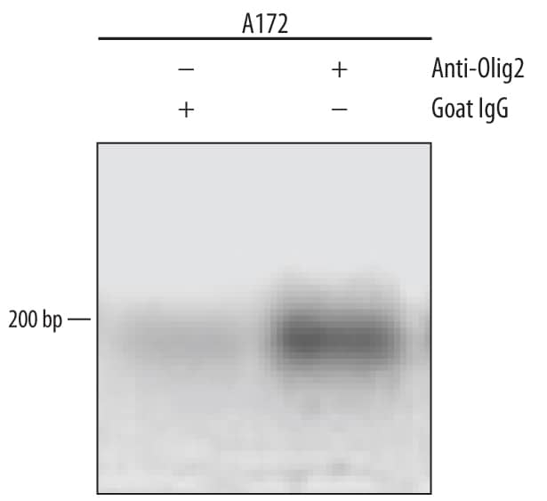
AB-108-C) for 15 minutes in an ultrasonic bath, followed by Biotinylated Anti-Goat IgG Secondary Antibody (Catalog #
BAF109). Immunocomplexes were captured using 50 µL of MagCellect Streptavidin Ferrofluid (Catalog #
MAG999) and DNA was purified using chelating resin solution. The
p21promoter was detected by standard PCR." class="big_thumb" />
VC004). Before incubation with the primary antibody, tissue was subjected to heat-induced epitope retrieval using Antigen Retrieval Reagent-Basic (
CTS013). Tissue was stained using DAB (brown) and counterstained with hematoxylin (blue). Specific staining was localized to cell nuclei. Staining was performed using our protocol for IHC Staining with VisUCyte HRP Polymer Detection Reagents." class="big_lightbox">
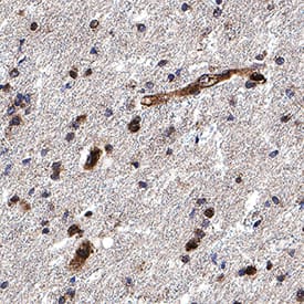
VC004). Before incubation with the primary antibody, tissue was subjected to heat-induced epitope retrieval using Antigen Retrieval Reagent-Basic (
CTS013). Tissue was stained using DAB (brown) and counterstained with hematoxylin (blue). Specific staining was localized to cell nuclei. Staining was performed using our protocol for IHC Staining with VisUCyte HRP Polymer Detection Reagents." class="big_thumb" />
VC004). Before incubation with the primary antibody, tissue was subjected to heat-induced epitope retrieval using Antigen Retrieval Reagent-Basic (
CTS013). Tissue was stained using DAB (brown) and counterstained with hematoxylin (blue). Specific staining was localized to cell nuclei. Staining was performed using our protocol for IHC Staining with VisUCyte HRP Polymer Detection Reagents." class="big_lightbox">
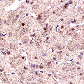
VC004). Before incubation with the primary antibody, tissue was subjected to heat-induced epitope retrieval using Antigen Retrieval Reagent-Basic (
CTS013). Tissue was stained using DAB (brown) and counterstained with hematoxylin (blue). Specific staining was localized to cell nuclei. Staining was performed using our protocol for IHC Staining with VisUCyte HRP Polymer Detection Reagents." class="big_thumb" />


AF2418) at 3ug/mL with overnight incubation at 4 degrees Celsius followed by incubation with anti-goat IgG VisUCyte HRP Polymer Antibody (Catalog #
VC004) and DAB chromogen (yellow-brown). Tissue was counterstained with hematoxylin (blue). Specific staining was localized to glial cells. " class="big_lightbox">

AF2418) at 3ug/mL with overnight incubation at 4 degrees Celsius followed by incubation with anti-goat IgG VisUCyte HRP Polymer Antibody (Catalog #
VC004) and DAB chromogen (yellow-brown). Tissue was counterstained with hematoxylin (blue). Specific staining was localized to glial cells. " class="big_thumb" />
 MAB1326). Cells were incubated with primary antibodies for 3 hours at room temperature. Cells were stained for Olig2 using the NorthernLights™ 637-conjugated Anti-Goat IgG Secondary Antibody (red; Catalog # NL002), and stained for O4 using an anti-mouse IgM secondary antibody (pseudo-stained green). View our protocol for Fluorescent ICC Staining of Cells on Coverslips." alt="Olig2 and Oligodendrocyte Marker O4 were detected in 7 day differentiated rat cortical stem cells using 10 µg/mL Goat Anti-Human/Mouse/Rat Olig2 Antigen Affinity-purified Polyclonal Antibody (Catalog # AF2418) and 10 µg/mL Goat Anti-Human/Mouse/Rat/Chicken O4 Monoclonal Antibody (Catalog # MAB1326). Cells were incubated with primary antibodies for 3 hours at room temperature. Cells were stained for Olig2 using the NorthernLights™ 637-conjugated Anti-Goat IgG Secondary Antibody (red; Catalog # NL002), and stained for O4 using an anti-mouse IgM secondary antibody (pseudo-stained green). View our protocol for Fluorescent ICC Staining of Cells on Coverslips." class="big_thumb" />


 AB-108-C) for 15 minutes in an ultrasonic bath, followed by Biotinylated Anti-Goat IgG Secondary Antibody (Catalog #
  CTS008) and counterstained with hematoxylin (blue). Specific staining was localized to developing brain. View our protocol for
  VC004). Before incubation with the primary antibody, tissue was subjected to heat-induced epitope retrieval using Antigen Retrieval Reagent-Basic (
 VC004). Before incubation with the primary antibody, tissue was subjected to heat-induced epitope retrieval using Antigen Retrieval Reagent-Basic (


 AF2418) at 3ug/mL with overnight incubation at 4 degrees Celsius followed by incubation with anti-goat IgG VisUCyte HRP Polymer Antibody (Catalog #
![Immunohistochemistry: Olig2 Antibody [NBP1-28667] - Mouse cerebral microslice section (coronal, P14) was co-immunostained with anti-Olig2 and anti-NeuN antibodies. Green: Olig2, Red: NeuN. Anti-Olig2 was used at a 1:800 dilution. This image was submitted via customer Review.](http://images.novusbio.com/fullsize/Olig2-Antibody-Immunohistochemistry-NBP1-28667-img0005.jpg "Immunohistochemistry: Olig2 Antibody [NBP1-28667] - Mouse cerebral microslice section (coronal, P14) was co-immunostained with anti-Olig2 and anti-NeuN antibodies. Green: Olig2, Red: NeuN. Anti-Olig2 was used at a 1:800 dilution. This image was submitted via customer Review.")
![<b>Biological Strategies Validation. </b>Immunocytochemistry/Immunofluorescence: Olig2 Antibody [NBP1-28667] - Rat cortical neuroepithelial cells with anti-Olig2 (red). The cells were treated with basic FGF for 30 hours prior to staining (this induces Olig2 expression in these primary cells).](http://images.novusbio.com/fullsize/Olig2-Antibody-Immunocytochemistry-Immunofluorescence-NBP1-28667-img0004.jpg)
![Immunohistochemistry-Frozen: Olig2 Antibody [NBP1-28667] - Mouse brain cortex. Antibody at 1:300. Purple: Olig2. Green: CD31. Image submitted by a verified customer review.](http://images.novusbio.com/fullsize/Olig2-Antibody-Immunohistochemistry-Frozen-NBP1-28667-img0006.jpg)

![Western Blot: Olig2 Antibody [NBP2-41269] - Western blot analysis of OLIG2 in EL4 cell lysate with OLIG2 antibody at 1 ug/ml in (A) the absence and (B) the presence of blocking peptide.](http://images.novusbio.com/fullsize/Olig2-Antibody-Western-Blot-NBP2-41269-img0001.jpg "Western Blot: Olig2 Antibody [NBP2-41269] - Western blot analysis of OLIG2 in EL4 cell lysate with OLIG2 antibody at 1 ug/ml in (A) the absence and (B) the presence of blocking peptide.")
![Immunohistochemistry: Olig2 Antibody [NBP2-41269] - Staining of OLIG2 in mouse brain tissue with OLIG2 Antibodyat 5 ug/mL.](http://images.novusbio.com/fullsize/Olig2-Antibody-Immunohistochemistry-NBP2-41269-img0005.jpg)
![Immunocytochemistry/ Immunofluorescence: Olig2 Antibody - BSA Free [NBP2-41269] - Immunofluorescence of Olig2 in mouse brain tissue with Olig2 Antibody at 20 ug/mL.<br><br>Green: Olig2 antibody <br> Red: Phylloidin staining<br> Blue: DAPI staining](http://images.novusbio.com/fullsize/nbp2-41269_rabbit-polyclonal-olig2-antibody-271120242045633.gif)
![Immunocytochemistry/ Immunofluorescence: Olig2 Antibody - BSA Free [NBP2-41269] - Immunofluorescence of Olig2 in rat brain tissue with Olig2 antibody at 20 ug/mL.](http://images.novusbio.com/fullsize/nbp2-41269_rabbit-polyclonal-olig2-antibody-2711202420285837.jpg)
![Immunohistochemistry: Olig2 Antibody - BSA Free [NBP2-41269] - Immunohistochemistry of Olig2 in rat brain tissue with Olig2 antibody at 5 ug/mL.](http://images.novusbio.com/fullsize/nbp2-41269_rabbit-polyclonal-olig2-antibody-2711202419432216.jpg)
![Western Blot: Olig2 Antibody (064) [NBP2-89201] - A: Jurkat Whole Cell Lysate Lysates/proteins at 30 ug per lane. SecondaryGoat Anti-Rabbit IgG H&L (Dylight800) at 1/10000 dilution. Developed using the Odyssey technique. Performed under reducing conditions. Predicted band size:32 kDa Observed band size:37 kDa](http://images.novusbio.com/fullsize/Olig2-Antibody-064-Western-Blot-NBP2-89201-img0003.jpg "Western Blot: Olig2 Antibody (064) [NBP2-89201] - A: Jurkat Whole Cell Lysate Lysates/proteins at 30 ug per lane. SecondaryGoat Anti-Rabbit IgG H&L (Dylight800) at 1/10000 dilution. Developed using the Odyssey technique. Performed under reducing conditions. Predicted band size:32 kDa Observed band size:37 kDa")
![Immunohistochemistry-Paraffin: Olig2 Antibody (064) [NBP2-89201] - Staining of human olig-2 in human brain with rabbit monoclonal antibody (1:200).](http://images.novusbio.com/fullsize/Olig2-Antibody-064-Immunohistochemistry-Paraffin-NBP2-89201-img0002.jpg)
![Immunohistochemistry-Paraffin: Olig2 Antibody (064) [NBP2-89201] - Staining of human olig-2 in human glioma with rabbit monoclonal antibody (1:200).](http://images.novusbio.com/fullsize/Olig2-Antibody-064-Immunohistochemistry-Paraffin-NBP2-89201-img0001.jpg)
![Flow Cytometry: Rabbit Monoclonal Olig2 Antibody (064) [NBP2-89201] - Mouse brain cells fixed and stained for Olig2 protein: 1:100. AF405 conjugated version of antibody used (Catalog # NBP2-89201AF405). Image from a verified customer review.](http://images.novusbio.com/fullsize/nbp2-89201_rabbit-monoclonal-olig2-antibody-064-flow-cytometry-1052024144135..png)
![Flow Cytometry: Rabbit Monoclonal Olig2 Antibody (064) [NBP2-89201] - Rat brain cells fixed and stained with Olig2. AF405 conjugated version of antibody used (Catalog # NBP2-89201AF405). Image from a verified customer review.](http://images.novusbio.com/fullsize/nbp2-89201_rabbit-monoclonal-olig2-antibody-064-flow-cytometry-1052024144528..png)
![Immunocytochemistry: Olig2 Antibody (3C9) [H00010215-M03] - Imaging of Optic Nerve: Red-H00010215-M03(Olig2), Blue-DAPI. This image was submitted via customer Review.](http://images.novusbio.com/fullsize/Olig2-Antibody-3C9-Immunocytochemistry-H00010215-M03-img0006.jpg "Immunocytochemistry: Olig2 Antibody (3C9) [H00010215-M03] - Imaging of Optic Nerve: Red-H00010215-M03(Olig2), Blue-DAPI. This image was submitted via customer Review.")
![Immunohistochemistry-Paraffin: Olig2 Antibody (3C9) [H00010215-M03] - Analysis of monoclonal antibody to OLIG2 on formalin-fixed paraffin-embedded human placenta. Antibody concentration 3 ug/ml](http://images.novusbio.com/fullsize/Olig2-Antibody-3C9-Immunohistochemistry-Paraffin-H00010215-M03-img0005.jpg)
![Immunocytochemistry/Immunofluorescence: Olig2 Antibody (3C9) [H00010215-M03] - Analysis of monoclonal antibody to OLIG2 on HeLa cell. Antibody concentration 10 ug/ml](http://images.novusbio.com/fullsize/Olig2-Antibody-3C9-Immunocytochemistry-Immunofluorescence-H00010215-M03-img0004.jpg)
![Western Blot: Olig2 Antibody [NBP2-27301] - Mouse brain lysate was separated on a 12% gel using SDS-PAGE, protein transferred to PVDF membrane and blocked in 5% milk-TBST. The membrane was probed with 2 ug/mL Olig2 antibody in 1% milk and detected with an anti-rabbit HRP conjugated secondary using chemiluminescence.](http://images.novusbio.com/fullsize/Olig2-Antibody-Western-Blot-NBP2-27301-img0003.jpg "Western Blot: Olig2 Antibody [NBP2-27301] - Mouse brain lysate was separated on a 12% gel using SDS-PAGE, protein transferred to PVDF membrane and blocked in 5% milk-TBST. The membrane was probed with 2 ug/mL Olig2 antibody in 1% milk and detected with an anti-rabbit HRP conjugated secondary using chemiluminescence.")
![Immunocytochemistry/Immunofluorescence: Olig2 Antibody [NBP2-27301] - Neuro2a cells were fixed for 10 minutes using 10% formalin and then permeabilized for 5 minutes using 1X TBS + 0.5% Triton X-100. The cells were incubated with Olig2 NBP2-27301 at a 1:200 dilution overnight at 4 degrees Celsius and detected using Dylight 488 (Green). Alpha tubulin was used as a co-stain at a 1:1000 dilution and detected with Dylight 550 (Red). Nuclei were detected with DAPI (Blue). Cells were imaged using a 40X objective.](http://images.novusbio.com/fullsize/Olig2-Antibody-Immunocytochemistry-Immunofluorescence-NBP2-27301-img0002.jpg)
![Immunohistochemistry-Paraffin: Olig2 Antibody [NBP2-27301] - OLIG2 Antibody [NBP2-27301] - FFPE human cerebellum probed with OLIG2 antibody at 1:200.](http://images.novusbio.com/fullsize/OLIG2-Antibody-Immunohistochemistry-Paraffin-NBP2-27301-img0001.jpg)
![Immunohistochemistry-Paraffin: Olig2 Antibody [NBP1-88634] - Staining in human cerebral cortex and liver tissues . Corresponding OLIG2 RNA-seq data are presented for the same tissues.](http://images.novusbio.com/fullsize/Olig2-Antibody-Immunohistochemistry-Paraffin-NBP1-88634-img0015.jpg "<b>Orthogonal Strategies Validation. </b>Immunohistochemistry-Paraffin: Olig2 Antibody [NBP1-88634] - Staining in human cerebral cortex and liver tissues . Corresponding OLIG2 RNA-seq data are presented for the same tissues.")
![<b>Orthogonal Strategies Validation. </b>Western Blot: Olig2 Antibody [NBP1-88634] - Analysis in human cerebral cortex tissue.](http://images.novusbio.com/fullsize/Olig2-Antibody-Western-Blot-NBP1-88634-img0016.jpg)
![Immunohistochemistry-Paraffin: Olig2 Antibody [NBP1-88634] - Staining of human endometrium shows no positivity in glandular cells as expected.](http://images.novusbio.com/fullsize/Olig2-Antibody-Immunohistochemistry-Paraffin-NBP1-88634-img0017.jpg)
![Immunohistochemistry-Paraffin: Olig2 Antibody [NBP1-88634] - Staining of human cerebral cortex shows moderate nuclear positivity in oligodendrocytes.](http://images.novusbio.com/fullsize/Olig2-Antibody-Immunohistochemistry-Paraffin-NBP1-88634-img0012.jpg)
![Immunohistochemistry-Paraffin: Olig2 Antibody [NBP1-88634] - Staining of human glioma shows moderate nuclear positivity in tumor cells.](http://images.novusbio.com/fullsize/Olig2-Antibody-Immunohistochemistry-Paraffin-NBP1-88634-img0013.jpg)
![Immunohistochemistry-Paraffin: Olig2 Antibody [NBP1-88634] - Staining of human liver shows no positivity as expected.](http://images.novusbio.com/fullsize/Olig2-Antibody-Immunohistochemistry-Paraffin-NBP1-88634-img0014.jpg)

![Immunocytochemistry/Immunofluorescence: Olig2 Antibody (HL1072) [NBP3-13712] - Olig2 antibody detects Olig2 protein at oligodendrocyte by immunofluorescent analysis. Sample: DIV10 rat cortical neuron-no AraC cells were fixed in 4% paraformaldehyde at RT for 15 min. Green: Olig2 stained by Olig2 antibody (NBP3-13712) diluted at 1:500. Red: Tau, a marker, stained by Tau antibody [GT287] diluted at 1:500.](http://images.novusbio.com/fullsize/Olig2-Antibody-HL1072-Immunocytochemistry-Immunofluorescence-NBP3-13712-img0002.jpg "Immunocytochemistry/Immunofluorescence: Olig2 Antibody (HL1072) [NBP3-13712] - Olig2 antibody detects Olig2 protein at oligodendrocyte by immunofluorescent analysis. Sample: DIV10 rat cortical neuron-no AraC cells were fixed in 4% paraformaldehyde at RT for 15 min. Green: Olig2 stained by Olig2 antibody (NBP3-13712) diluted at 1:500. Red: Tau, a marker, stained by Tau antibody [GT287] diluted at 1:500.")
![Immunohistochemistry-Frozen: Olig2 Antibody (HL1072) [NBP3-13712] - Olig2 antibody [HL1072] detects Olig2 protein at nucleus by immunohistochemical analysis. Sample: Frozen-sectioned mouse brain. Green: Olig2 stained by Olig2 antibody [HL1072] (NBP3-13712) diluted at 1:100. Blue: Fluoroshield with DAPI. Antigen Retrieval: ice-cold MeOH for 5 min](http://images.novusbio.com/fullsize/Olig2-Antibody-HL1072-Immunohistochemistry-Frozen-NBP3-13712-img0001.jpg)
![Immunohistochemistry-Paraffin: Olig2 Antibody (HL1072) [NBP3-13712] - Olig2 antibody [HL1072] detects Olig2 protein at nucleus by immunohistochemical analysis. Sample: Paraffin-embedded mouse E10.5 embryo. Green: Olig2 stained by Olig2 antibody [HL1072] (NBP3-13712) diluted at 1:200. Red: beta Tubulin 3/ Tuj1, a nerual marker, stained by beta Tubulin 3/ Tuj1 antibody [GT11710] (NBP2-43559) diluted at 1:. Blue: Fluoroshield with DAPI. Antigen Retrieval: Citrate buffer, pH 6.0, 15 min](http://images.novusbio.com/fullsize/Olig2-Antibody-HL1072-Immunohistochemistry-Paraffin-NBP3-13712-img0004.jpg)
![Immunohistochemistry-Paraffin: Olig2 Antibody (HL1072) [NBP3-13712] - Olig2 antibody [HL1072] detects Olig2 protein at nucleus by immunohistochemical analysis.</br>Sample: Paraffin-embedded mouse brain.</br>Olig2 stained by Olig2 antibody [HL1072] (NBP3-13712) diluted at 1:100.</br>Antigen Retrieval: Citrate buffer, pH 6.0, 15 min](http://images.novusbio.com/fullsize/nbp3-13712_rabbit-olig2-mab-hl1072-91020242014688.jpg)
![Western Blot: Olig2 Antibody (1G11) [NBP2-37636] - Western blot analysis using OLIG2 mAb against HEK293 (1) and OLIG2(AA: 1-122)-hIgGFc transfected HEK293 (2) cell lysate.](http://images.novusbio.com/fullsize/Olig2-Antibody-1G11-Western-Blot-NBP2-37636-img0002.jpg "Western Blot: Olig2 Antibody (1G11) [NBP2-37636] - Western blot analysis using OLIG2 mAb against HEK293 (1) and OLIG2(AA: 1-122)-hIgGFc transfected HEK293 (2) cell lysate.")
![Immunocytochemistry/Immunofluorescence: Olig2 Antibody (1G11) [NBP2-37636] - Immunofluorescence analysis of U251 cells using OLIG2 mouse mAb (green). Blue: DRAQ5 fluorescent DNA dye.](http://images.novusbio.com/fullsize/Olig2-Antibody-1G11-Immunocytochemistry-Immunofluorescence-NBP2-37636-img0003.jpg)
![Immunohistochemistry: Olig2 Antibody (1G11) [NBP2-37636] - Immunohistochemical analysis of paraffin-embedded muscle tissues using OLIG2 mouse mAb with DAB staining.](http://images.novusbio.com/fullsize/Olig2-Antibody-1G11-Immunohistochemistry-NBP2-37636-img0004.jpg)
![ELISA: Olig2 Antibody (1G11) [NBP2-37636] - Red: Control Antigen (100ng); Purple: Antigen (10ng); Green: Antigen (50ng); Blue: Antigen (100ng);](http://images.novusbio.com/fullsize/Olig2-Antibody-1G11-ELISA-NBP2-37636-img0001.jpg)
![Immunohistochemistry-Paraffin: Olig2 Antibody (OLIG2/6695R) [NBP3-13830] - Formalin-fixed, paraffin-embedded human glioma stained withOlig2 antibody (OLIG2/6695R).](http://images.novusbio.com/fullsize/Olig2-Antibody-OLIG2-6695R-Immunohistochemistry-Paraffin-NBP3-13830-img0004.jpg "Immunohistochemistry-Paraffin: Olig2 Antibody (OLIG2/6695R) [NBP3-13830] - Formalin-fixed, paraffin-embedded human glioma stained withOlig2 antibody (OLIG2/6695R).")
![Immunohistochemistry-Paraffin: Olig2 Antibody (OLIG2/6695R) [NBP3-13830] - Formalin-fixed, paraffin-embedded human cerebrum stained with Olig2 antibody (OLIG2/6695R).](http://images.novusbio.com/fullsize/Olig2-Antibody-OLIG2-6695R-Immunohistochemistry-Paraffin-NBP3-13830-img0001.jpg)
![Immunohistochemistry-Paraffin: Olig2 Antibody (OLIG2/6695R) [NBP3-13830] - Formalin-fixed, paraffin-embedded human oligodendroglioma stained with Olig2 antibody (OLIG2/6695R).](http://images.novusbio.com/fullsize/Olig2-Antibody-OLIG2-6695R-Immunohistochemistry-Paraffin-NBP3-13830-img0002.jpg)
![Immunohistochemistry-Paraffin: Olig2 Antibody (OLIG2/6695R) [NBP3-13830] - Formalin-fixed, paraffin-embedded human cerebellum stained with Olig2 antibody (OLIG2/6695R).](http://images.novusbio.com/fullsize/Olig2-Antibody-OLIG2-6695R-Immunohistochemistry-Paraffin-NBP3-13830-img0003.jpg)
![Immunohistochemistry-Paraffin: Olig2 Antibody (OLIG2/6695R) [NBP3-13830] - IHC analysis of formalin-fixed, paraffin-embedded human brain. Stained with Olig2 antibody (OLIG2/6695R) at 2ug/ml in PBS for 30min RT. Inset: PBS instead of primary; secondary antibody negative control.](http://images.novusbio.com/fullsize/Olig2-Antibody-OLIG2-6695R-Immunohistochemistry-Paraffin-NBP3-13830-img0005.jpg)


