HAF017). For additional reference, recombinant human 14-3-3 beta, epsilon, eta, gamma, sigma, theta, and zeta (2 ng/lane) were included. A specific band for 14-3-3 sigma was detected at approximately 28 kDa (as indicated). This experiment was conducted under reducing conditions and using
Immunoblot Buffer Group 1." class="big_lightbox">

HAF017). For additional reference, recombinant human 14-3-3 beta, epsilon, eta, gamma, sigma, theta, and zeta (2 ng/lane) were included. A specific band for 14-3-3 sigma was detected at approximately 28 kDa (as indicated). This experiment was conducted under reducing conditions and using
Immunoblot Buffer Group 1." alt="Western blot shows lysates of A431 human epithelial carcinoma cell line and mouse brain tissue. PVDF membrane was probed with 1 µg/mL Goat Anti-Human/ Mouse/Rat 14-3-3s Antigen Affinity-purified Polyclonal Antibody (Catalog # AF4424) followed by HRP-conjugated Anti-Goat IgG Secondary Antibody (Catalog #
HAF017). For additional reference, recombinant human 14-3-3 beta, epsilon, eta, gamma, sigma, theta, and zeta (2 ng/lane) were included. A specific band for 14-3-3 sigma was detected at approximately 28 kDa (as indicated). This experiment was conducted under reducing conditions and using
Immunoblot Buffer Group 1." class="big_thumb" />
HAF017). A specific band was detected for 14-3-3s at approximately 28 kDa (as indicated) in the parental HeLa cell line, but is not detectable in knockout HeLa cell line. GAPDH (Catalog #
AF5718) is shown as a loading control. This experiment was conducted under reducing conditions and using
Immunoblot Buffer Group 1." class="big_lightbox">
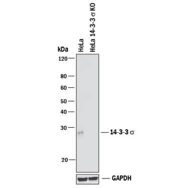
HAF017). A specific band was detected for 14-3-3s at approximately 28 kDa (as indicated) in the parental HeLa cell line, but is not detectable in knockout HeLa cell line. GAPDH (Catalog #
AF5718) is shown as a loading control. This experiment was conducted under reducing conditions and using
Immunoblot Buffer Group 1." class="big_thumb" />
 HAF017). For additional reference, recombinant human 14-3-3 beta, epsilon, eta, gamma, sigma, theta, and zeta (2 ng/lane) were included. A specific band for 14-3-3 sigma was detected at approximately 28 kDa (as indicated). This experiment was conducted under reducing conditions and using Immunoblot Buffer Group 1." alt="Western blot shows lysates of A431 human epithelial carcinoma cell line and mouse brain tissue. PVDF membrane was probed with 1 µg/mL Goat Anti-Human/ Mouse/Rat 14-3-3s Antigen Affinity-purified Polyclonal Antibody (Catalog # AF4424) followed by HRP-conjugated Anti-Goat IgG Secondary Antibody (Catalog # HAF017). For additional reference, recombinant human 14-3-3 beta, epsilon, eta, gamma, sigma, theta, and zeta (2 ng/lane) were included. A specific band for 14-3-3 sigma was detected at approximately 28 kDa (as indicated). This experiment was conducted under reducing conditions and using Immunoblot Buffer Group 1." class="big_thumb" />


![Western Blot: 14-3-3 sigma/Stratifin Antibody [NBP2-27072] - Analysis of 14-3-3 sigma (169 amino acid isoform) using 14-3-3 sigma antibody. Human brain tissue in the 1) absence and 2) presence of immunizing peptide, 3) mouse brain and 4) rat brain tissue probed with 14-3-3 sigma antibody at 3 ug/ml.](http://images.novusbio.com/fullsize/14-3-3-sigma-Stratifin-Antibody-Western-Blot-NBP2-27072-img0004.jpg "Western Blot: 14-3-3 sigma/Stratifin Antibody [NBP2-27072] - Analysis of 14-3-3 sigma (169 amino acid isoform) using 14-3-3 sigma antibody. Human brain tissue in the 1) absence and 2) presence of immunizing peptide, 3) mouse brain and 4) rat brain tissue probed with 14-3-3 sigma antibody at 3 ug/ml.")
![Immunohistochemistry-Paraffin: 14-3-3 sigma/Stratifin Antibody [NBP2-27072] - Formalin-fixed, paraffin-embedded human tonsil (squamous epithelium) probed with 14-3-3 sigma antibody at 10 ug/ml.](http://images.novusbio.com/fullsize/14-3-3-sigma-Stratifin-Antibody-Immunohistochemistry-Paraffin-NBP2-27072-img0005.jpg)
![Western Blot: 14-3-3 sigma/Stratifin Antibody [NBP2-27072] - analysis of 14-3-3 sigma (248 amino acid isoform) using 14-3-3 sigma polyclonal antibody. Human A431 cell lysate in the 1) absence and 2) presence of immunizing peptide, and 3) HeLa cell lysate probed with 14-3-3 sigma antibody at 3 ug/ml.](http://images.novusbio.com/fullsize/14-3-3-sigma-Stratifin-Antibody-Western-Blot-NBP2-27072-img0002.jpg)
![Immunohistochemistry-Paraffin: 14-3-3 sigma/Stratifin Antibody [NBP1-80610] - Staining in human skin and skeletal muscle tissues using anti-SFN antibody. Corresponding SFN RNA-seq data are presented for the same tissues.](http://images.novusbio.com/fullsize/14-3-3-sigma-Stratifin-Antibody-Immunohistochemistry-Paraffin-NBP1-80610-img0012.jpg "<b>Orthogonal Strategies Validation. </b>Immunohistochemistry-Paraffin: 14-3-3 sigma/Stratifin Antibody [NBP1-80610] - Staining in human skin and skeletal muscle tissues using anti-SFN antibody. Corresponding SFN RNA-seq data are presented for the same tissues.")
![<b>Orthogonal Strategies Validation. </b>Western Blot: 14-3-3 sigma/Stratifin Antibody [NBP1-80610] - Analysis in human cell lines A-431 and SK-MEL-30. Corresponding RNA-seq data are presented for the same cell lines. Loading control: Anti-HSP90B1.](http://images.novusbio.com/fullsize/14-3-3-sigma-Stratifin-Antibody-Western-Blot-NBP1-80610-img0010.jpg)
![Western Blot: 14-3-3 sigma/Stratifin Antibody [NBP1-80610] - Lane 1: Marker [kDa] 230, 130, 95, 72, 56, 36, 28, 17, 11. Lane 2: Human cell line RT-4. Lane 3: Human cell line U-251MG sp. Lane 4: Human plasma (IgG/HSA depleted). Lane 5: Human liver tissue. Lane 6: Human tonsil tissue](http://images.novusbio.com/fullsize/14-3-3-sigma-Stratifin-Antibody-Western-Blot-NBP1-80610-img0006.jpg)
![Western Blot: 14-3-3 sigma/Stratifin Antibody [NBP1-80610] - Lane 1: NIH-3T3 cell lysate (Mouse embryonic fibroblast cells). Lane 2: NBT-II cell lysate (Rat Wistar bladder tumor cells).](http://images.novusbio.com/fullsize/14-3-3-sigma-Stratifin-Antibody-Western-Blot-NBP1-80610-img0007.jpg)
![Immunohistochemistry-Paraffin: 14-3-3 sigma/Stratifin Antibody [NBP1-80610] - Staining of human skin shows strong cytoplasmic positivity in epidermal cells.](http://images.novusbio.com/fullsize/14-3-3-sigma-Stratifin-Antibody-Immunohistochemistry-Paraffin-NBP1-80610-img0002.jpg)
![Immunohistochemistry-Paraffin: 14-3-3 sigma/Stratifin Antibody [NBP1-80610] - Staining of human skeletal muscle shows low expression as expected.](http://images.novusbio.com/fullsize/14-3-3-sigma-Stratifin-Antibody-Immunohistochemistry-Paraffin-NBP1-80610-img0011.jpg)
 HAF017). A specific band was detected for 14-3-3s at approximately 28 kDa (as indicated) in the parental HeLa cell line, but is not detectable in knockout HeLa cell line. GAPDH (Catalog #
 ![Western Blot: 14-3-3 sigma/Stratifin Antibody [NB100-2465] - Staining of Mouse (A) and Rat (B) Skin lysate (35 ug protein in RIPA buffer). Antibody at 0.03 ug/mL. Detected by chemiluminescence.](http://images.novusbio.com/fullsize/14-3-3-sigma-Stratifin-Antibody-Western-Blot-NB100-2465-img0007.jpg "Western Blot: 14-3-3 sigma/Stratifin Antibody [NB100-2465] - Staining of Mouse (A) and Rat (B) Skin lysate (35 ug protein in RIPA buffer). Antibody at 0.03 ug/mL. Detected by chemiluminescence.")
![Immunohistochemistry-Paraffin: 14-3-3 sigma/Stratifin Antibody [NB100-2465] - Staining of paraffin embedded human skin. Antibody at 2 ug/mL. Steamed antigen retrieval with Tris/EDTA buffer pH 9, HRP-staining. Image shows strong nuclear staining in selected cells of the basal layer.](http://images.novusbio.com/fullsize/14-3-3-sigma-Stratifin-Antibody-Immunohistochemistry-Paraffin-NB100-2465-img0004.jpg)
![Western Blot: 14-3-3 sigma/Stratifin Antibody [NB100-2465] - (0.1ug/ml) staining of human skin lysate (35ug protein in RIPA buffer). Primary incubation was 1 hour. Detected by chemiluminescence.](http://images.novusbio.com/fullsize/14-3-3-sigma-Stratifin-Antibody-Western-Blot-NB100-2465-img0003.jpg)
![Western Blot: 14-3-3 sigma/Stratifin Antibody [NB100-2465] - Staining of human skin lysate (35 ug protein in RIPA buffer). Antibody at 0.03 ug/mL. Primary incubation was 1 hour. Detected by chemiluminescence.](http://images.novusbio.com/fullsize/14-3-3-sigma-Stratifin-Antibody-Western-Blot-NB100-2465-img0005.jpg)
![Western Blot: 14-3-3 sigma/Stratifin Antibody [NB100-2465] - Staining of A431 (A) and U2OS (B) cell lysate (35 ug protein in RIPA buffer). Antibody at 0.03 ug/mL. Detected by chemiluminescence.](http://images.novusbio.com/fullsize/14-3-3-sigma-Stratifin-Antibody-Western-Blot-NB100-2465-img0006.jpg)
![Western Blot: 14-3-3 sigma/Stratifin Antibody [NBP2-98309] - Anti-14-3-3 sigma/Stratifin rabbit polyclonal antibody at 1:500 dilution. Lane A: NIH3T3 Whole Cell Lysate. Lane B: Raw 264.7 Whole Cell Lysate. Lysates/proteins at 30 ug per lane. Secondary Goat Anti-Rabbit IgG (H+L)/HRP at 1/10000 dilution. Developed using the ECL technique. Performed under reducing conditions. Predicted band size: 27 kDa](http://images.novusbio.com/fullsize/14-3-3-sigma-Stratifin-Antibody-Western-Blot-NBP2-98309-img0006.jpg "Western Blot: 14-3-3 sigma/Stratifin Antibody [NBP2-98309] - Anti-14-3-3 sigma/Stratifin rabbit polyclonal antibody at 1:500 dilution. Lane A: NIH3T3 Whole Cell Lysate. Lane B: Raw 264.7 Whole Cell Lysate. Lysates/proteins at 30 ug per lane. Secondary Goat Anti-Rabbit IgG (H+L)/HRP at 1/10000 dilution. Developed using the ECL technique. Performed under reducing conditions. Predicted band size: 27 kDa")
![Immunohistochemistry-Paraffin: 14-3-3 sigma/Stratifin Antibody [NBP2-98309] - Immunochemical staining of mouse 14-3-3 sigma/Stratifin in mouse skin with rabbit polyclonal antibody at 1:20000 dilution, formalin-fixed paraffin embedded sections.](http://images.novusbio.com/fullsize/14-3-3-sigma-Stratifin-Antibody-Immunohistochemistry-Paraffin-NBP2-98309-img0004.jpg)
![Immunoprecipitation: 14-3-3 sigma/Stratifin Antibody [NBP2-98309] - Mouse 14-3-3 sigma/Stratifin was immunoprecipitated using: Lane A: 0.5 mg NIH3T3 Whole Cell Lysate. 4 ul anti-Mouse 14-3-3 sigma/Stratifin rabbit polyclonal antibody and 60 ug of Immunomagnetic beads. Protein A/G. Primary antibody: Anti-Mouse 14-3-3 sigma/Stratifin rabbit polyclonal antibody, at 1:100 dilution. Secondary antibody: Goat Anti-Rabbit IgG (H+L)/HRP at 1/10000 dilution. Developed using the ECL technique. Performed under reducing conditions. Predicted band size: 30 kDa. Observed band size: 30 kDa](http://images.novusbio.com/fullsize/14-3-3-sigma-Stratifin-Antibody-Immunoprecipitation-NBP2-98309-img0005.jpg)
![Immunocytochemistry/Immunofluorescence: 14-3-3 sigma/Stratifin Antibody [NBP2-98309] - Immunofluorescence staining of 14-3-3 sigma/Stratifin in Raw264.7 cells. Cells were fixed with 4% PFA, permeabilzed with 0.1% Triton X-100 in PBS, blocked with 10% serum, and incubated with NBP2-98309 antibody (dilution ratio 1:200) at 4C overnight. Then cells were stained with the Alexa Fluor(R)488-conjugated Goat Anti-rabbit IgG Secondary antibody (green). Positive staining was localized to Cytoplasm.](http://images.novusbio.com/fullsize/nbp2-98309_rabbit-polyclonal-14-3-3-sigma-stratifin-antibody-23520252135137.png)
![Western Blot: 14-3-3 sigma/Stratifin Antibody (CPTC-SFN-2) [NBP3-07326] - Western Blot Analysis of Human PC-3 cell lysate using 14-3-3 sigma/Stratifin Mouse Monoclonal Antibody (CPTC-SFN-2).](http://images.novusbio.com/fullsize/14-3-3-sigma-Stratifin-Antibody-CPTC-SFN-2-Western-Blot-NBP3-07326-img0003.jpg "Western Blot: 14-3-3 sigma/Stratifin Antibody (CPTC-SFN-2) [NBP3-07326] - Western Blot Analysis of Human PC-3 cell lysate using 14-3-3 sigma/Stratifin Mouse Monoclonal Antibody (CPTC-SFN-2).")
![Immunohistochemistry-Paraffin: 14-3-3 sigma/Stratifin Antibody (CPTC-SFN-2) [NBP3-07326] - Formalin-fixed, paraffin-embedded human Skin stained with 14-3-3 sigma/Stratifin Mouse Monoclonal Antibody (CPTC-SFN-2).](http://images.novusbio.com/fullsize/14-3-3-sigma-Stratifin-Antibody-CPTC-SFN-2-Immunohistochemistry-Paraffin-NBP3-07326-img0002.jpg)
![Protein Array: 14-3-3 sigma/Stratifin Antibody (CPTC-SFN-2) [NBP3-07326] - Analysis of Protein Array containing more than 19,000 full-length human proteins using 14-3-3 sigma/Stratifin Mouse Monoclonal Antibody (CPTC-SFN-2).](http://images.novusbio.com/fullsize/14-3-3-sigma-Stratifin-Antibody-CPTC-SFN-2-Protein-Array-NBP3-07326-img0001.jpg)
![Western Blot: 14-3-3 sigma/Stratifin Antibody (8C8A9) [NBP3-16395] - Western blot analysis of extracts of various cell lines, using 14-3-3 sigma/Stratifin Rabbit mAb (NBP3-16395) at 1:1000 dilution. Secondary antibody: HRP Goat Anti-Rabbit IgG (H+L) at 1:10000 dilution. Lysates/proteins: 25ug per lane. Blocking buffer: 3% nonfat dry milk in TBST. Detection: ECL Basic Kit. Exposure time: 3min.](http://images.novusbio.com/fullsize/14-3-3-sigma-Stratifin-Antibody-8C8A9-Western-Blot-NBP3-16395-img0002.jpg "Western Blot: 14-3-3 sigma/Stratifin Antibody (8C8A9) [NBP3-16395] - Western blot analysis of extracts of various cell lines, using 14-3-3 sigma/Stratifin Rabbit mAb (NBP3-16395) at 1:1000 dilution. Secondary antibody: HRP Goat Anti-Rabbit IgG (H+L) at 1:10000 dilution. Lysates/proteins: 25ug per lane. Blocking buffer: 3% nonfat dry milk in TBST. Detection: ECL Basic Kit. Exposure time: 3min.")
![Immunohistochemistry-Paraffin: 14-3-3 sigma/Stratifin Antibody (8C8A9) [NBP3-16395] - Immunohistochemistry of paraffin-embedded human colon using 14-3-3 sigma/Stratifin Rabbit mAb (NBP3-16395) at dilution of 1:100 (40x lens).Perform microwave antigen retrieval with 10 mM PBS buffer pH 7.2 before commencing with IHC staining protocol.](http://images.novusbio.com/fullsize/14-3-3-sigma-Stratifin-Antibody-8C8A9-Immunohistochemistry-Paraffin-NBP3-16395-img0001.jpg)
![Western Blot: 14-3-3 sigma/Stratifin Overexpression Lysate (Adult Normal) [NBL1-15882] Left-Empty vector transfected control cell lysate (HEK293 cell lysate); Right -Over-expression Lysate for 14-3-3 sigma/Stratifin.](http://images.novusbio.com/fullsize/14-3-3-sigma-Stratifin-Overexpression-Lysate-Adult-Normal-Western-Blot-NBL1-15882-img0002.jpg "Western Blot: 14-3-3 sigma/Stratifin Overexpression Lysate (Adult Normal) [NBL1-15882] Left-Empty vector transfected control cell lysate (HEK293 cell lysate); Right -Over-expression Lysate for 14-3-3 sigma/Stratifin.")
![SDS-Page: Recombinant Human 14-3-3 sigma/Stratifin Protein [NBP1-30172] - 15% SDS-PAGE (3ug)](http://images.novusbio.com/fullsize/Recombinant-Human-14-3-3-sigma-Stratifin-Protein-SDS-Page-NBP1-30172-img0002.jpg "SDS-Page: Recombinant Human 14-3-3 sigma/Stratifin Protein [NBP1-30172] - 15% SDS-PAGE (3ug)")
![ELISA: Human 14-3-3 sigma/Stratifin - Ready-To-Use ELISA Kit (Colorimetric) [NBP3-40129] - Standard Curve Reference](http://images.novusbio.com/fullsize/nbp3-40129_human-14-3-3-sigma-stratifin-ready-to-use-elisa-kit-2110202415295678.png "ELISA: Human 14-3-3 sigma/Stratifin - Ready-To-Use ELISA Kit (Colorimetric) [NBP3-40129] - Standard Curve Reference")
![ELISA: Human 14-3-3 sigma/Stratifin ELISA Kit (Colorimetric) [NBP3-40130] - Standard Curve Reference](http://images.novusbio.com/fullsize/nbp3-40130_human-14-3-3-sigma-stratifin-elisa-kit-colorimetric-2110202415295649.png "ELISA: Human 14-3-3 sigma/Stratifin ELISA Kit (Colorimetric) [NBP3-40130] - Standard Curve Reference")

