MAB3286, Mouse IgG
2Bisotype), anti-HSP60 (
MAB1800, Mouse IgG
2Bisotype), anti-GAPDH (
MAB5718,Mouse IgG
1isotype), anti-Peroxiredoxin 6 (
MAB3490, Mouse IgG
3isotype), and anti-SOD1 (
MAB3418, Mouse IgG
2Aisotype). The left panel was then probed with 0.5 µg/mL of Rabbit Anti-Mouse IgG
2B(Catalog # MAB9636) followed by HRP-conjugated Anti-Rabbit IgG Secondary Antibody (
HAF008). Specific bands were detected for IgG
2Bisotype antibodies at approximately 60 and 90 kDa (as indicated). The right panel was probed with HRP-conjugated Anti-Mouse IgG Secondary Antibody (
HAF018) but not Rabbit Anti-Mouse IgG
2BMonoclonal Antibody (Catalog # MAB9636). This experiment was conducted under reducing conditions and using
Immunoblot Buffer Group 1." class="big_lightbox">
MAB3286, Mouse IgG
2Bisotype), anti-HSP60 (
MAB1800, Mouse IgG
2Bisotype), anti-GAPDH (
MAB5718,Mouse IgG
1isotype), anti-Peroxiredoxin 6 (
MAB3490, Mouse IgG
3isotype), and anti-SOD1 (
MAB3418, Mouse IgG
2Aisotype). The left panel was then probed with 0.5 µg/mL of Rabbit Anti-Mouse IgG
2B(Catalog # MAB9636) followed by HRP-conjugated Anti-Rabbit IgG Secondary Antibody (
HAF008). Specific bands were detected for IgG
2Bisotype antibodies at approximately 60 and 90 kDa (as indicated). The right panel was probed with HRP-conjugated Anti-Mouse IgG Secondary Antibody (
HAF018) but not Rabbit Anti-Mouse IgG
2BMonoclonal Antibody (Catalog # MAB9636). This experiment was conducted under reducing conditions and using
Immunoblot Buffer Group 1." alt="Western blot shows lysates of HepG2 and Jurkat cell lines. PVDF membrane was probed with anti-HSP90 (
MAB3286, Mouse IgG
2Bisotype), anti-HSP60 (
MAB1800, Mouse IgG
2Bisotype), anti-GAPDH (
MAB5718,Mouse IgG
1isotype), anti-Peroxiredoxin 6 (
MAB3490, Mouse IgG
3isotype), and anti-SOD1 (
MAB3418, Mouse IgG
2Aisotype). The left panel was then probed with 0.5 µg/mL of Rabbit Anti-Mouse IgG
2B(Catalog # MAB9636) followed by HRP-conjugated Anti-Rabbit IgG Secondary Antibody (
HAF008). Specific bands were detected for IgG
2Bisotype antibodies at approximately 60 and 90 kDa (as indicated). The right panel was probed with HRP-conjugated Anti-Mouse IgG Secondary Antibody (
HAF018) but not Rabbit Anti-Mouse IgG
2BMonoclonal Antibody (Catalog # MAB9636). This experiment was conducted under reducing conditions and using
Immunoblot Buffer Group 1." class="big_thumb" />
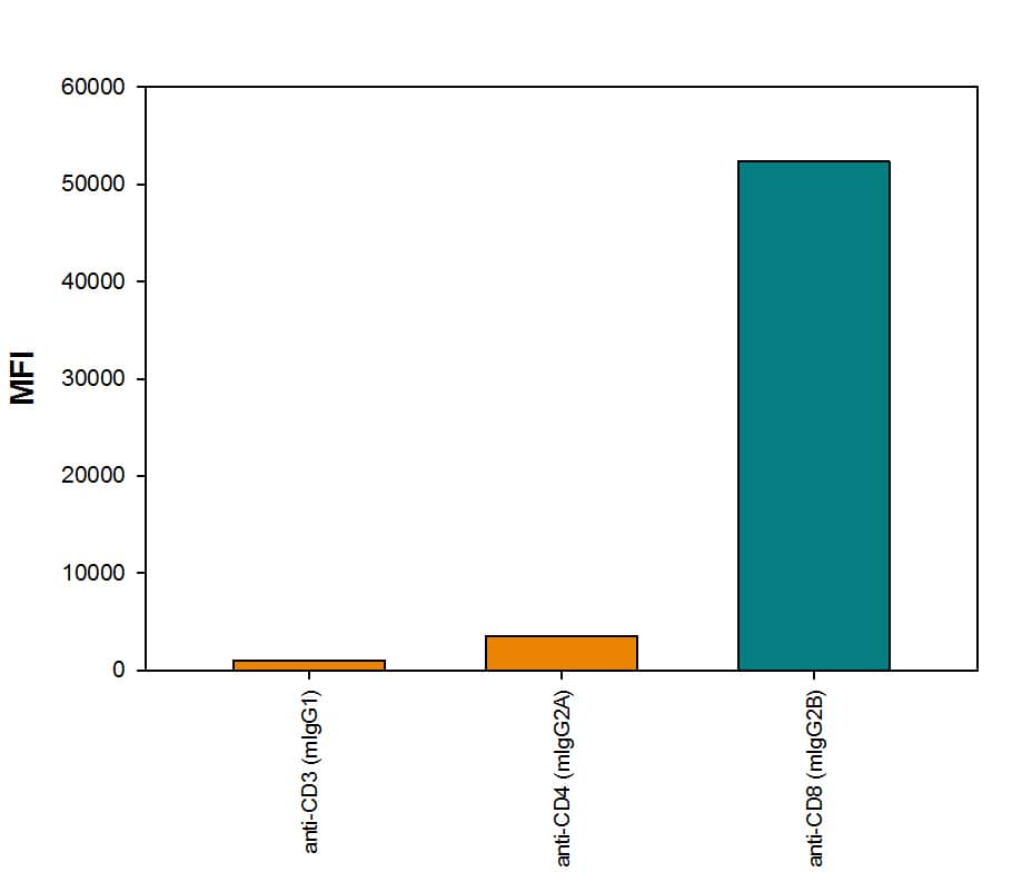
MAB100, Mouse IgG
1isotype), Mouse anti-Human CD4 Monoclonal Antibody (
MAB3791, Mouse IgG
2Aisotype), or Mouse anti-Human CD8a Monoclonal Antibody (
MAB1509, Mouse IgG
2Bisotype), followed by Rabbit anti-Mouse IgG
2B-specific secondary antibody (Catalog # MAB9636) then PE-conjugated Goat anti-Rabbit secondary antibody (
F0110). View our protocol for
Staining Membrane-associated Proteins." class="big_lightbox">
MAB100, Mouse IgG
1isotype), Mouse anti-Human CD4 Monoclonal Antibody (
MAB3791, Mouse IgG
2Aisotype), or Mouse anti-Human CD8a Monoclonal Antibody (
MAB1509, Mouse IgG
2Bisotype), followed by Rabbit anti-Mouse IgG
2B-specific secondary antibody (Catalog # MAB9636) then PE-conjugated Goat anti-Rabbit secondary antibody (
F0110). View our protocol for
Staining Membrane-associated Proteins." class="big_thumb" />
MAB859, Mouse IgG
1 isotype; aqua histogram), Mouse anti-Human MICA Monoclonal Antibody (Catalog #
MAB13001, Mouse IgG
2A isotype; dark blue histogram), or Mouse anti-Human MICA Monoclonal Antibody (Catalog #
MAB1300, Mouse IgG
2B isotype; orange histogram), followed by Rabbit anti-Mouse IgG
2B-specific secondary antibody (Catalog # MAB9636) then APC-conjugated Goat anti-Rabbit secondary antibody (Catalog #
F0101B). View our protocol for
Staining Membrane-associated Proteins." class="big_lightbox">

MAB859, Mouse IgG
1 isotype; aqua histogram), Mouse anti-Human MICA Monoclonal Antibody (Catalog #
MAB13001, Mouse IgG
2A isotype; dark blue histogram), or Mouse anti-Human MICA Monoclonal Antibody (Catalog #
MAB1300, Mouse IgG
2B isotype; orange histogram), followed by Rabbit anti-Mouse IgG
2B-specific secondary antibody (Catalog # MAB9636) then APC-conjugated Goat anti-Rabbit secondary antibody (Catalog #
F0101B). View our protocol for
Staining Membrane-associated Proteins." class="big_thumb" />
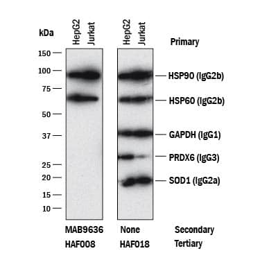
 MAB3286, Mouse IgG2Bisotype), anti-HSP60 (MAB1800, Mouse IgG2Bisotype), anti-GAPDH (MAB5718,Mouse IgG1isotype), anti-Peroxiredoxin 6 (MAB3490, Mouse IgG3isotype), and anti-SOD1 (MAB3418, Mouse IgG2Aisotype). The left panel was then probed with 0.5 µg/mL of Rabbit Anti-Mouse IgG2B(Catalog # MAB9636) followed by HRP-conjugated Anti-Rabbit IgG Secondary Antibody (HAF008). Specific bands were detected for IgG2Bisotype antibodies at approximately 60 and 90 kDa (as indicated). The right panel was probed with HRP-conjugated Anti-Mouse IgG Secondary Antibody (HAF018) but not Rabbit Anti-Mouse IgG2BMonoclonal Antibody (Catalog # MAB9636). This experiment was conducted under reducing conditions and using Immunoblot Buffer Group 1." alt="Western blot shows lysates of HepG2 and Jurkat cell lines. PVDF membrane was probed with anti-HSP90 (MAB3286, Mouse IgG2Bisotype), anti-HSP60 (MAB1800, Mouse IgG2Bisotype), anti-GAPDH (MAB5718,Mouse IgG1isotype), anti-Peroxiredoxin 6 (MAB3490, Mouse IgG3isotype), and anti-SOD1 (MAB3418, Mouse IgG2Aisotype). The left panel was then probed with 0.5 µg/mL of Rabbit Anti-Mouse IgG2B(Catalog # MAB9636) followed by HRP-conjugated Anti-Rabbit IgG Secondary Antibody (HAF008). Specific bands were detected for IgG2Bisotype antibodies at approximately 60 and 90 kDa (as indicated). The right panel was probed with HRP-conjugated Anti-Mouse IgG Secondary Antibody (HAF018) but not Rabbit Anti-Mouse IgG2BMonoclonal Antibody (Catalog # MAB9636). This experiment was conducted under reducing conditions and using Immunoblot Buffer Group 1." class="big_thumb" />


![Western Blot: IgG2b Antibody (RM108) [Unconjugated] [NBP2-62009] - Western blot of nonreduced(-) and reduced(+) mouse IgG2b, using NBP2-62009. This antibody reacts to nonreduced IgG2b (~150 kDa) much stronger than the reduced g2b form (~50 kDa).](http://images.novusbio.com/fullsize/IgG2b-Antibody-RM108-Unconjugated-Western-Blot-NBP2-62009-img0004.jpg "Western Blot: IgG2b Antibody (RM108) [Unconjugated] [NBP2-62009] - Western blot of nonreduced(-) and reduced(+) mouse IgG2b, using NBP2-62009. This antibody reacts to nonreduced IgG2b (~150 kDa) much stronger than the reduced g2b form (~50 kDa).")
![ELISA: IgG2b Antibody (RM108) - Azide and BSA Free [NBP2-62009] - A titer ELISA of mouse IgG2b. The plate was coated with different amounts of mouse IgG2b. A serial dilution of RM108 was used as the primary antibody. An alkaline phosphatase conjugated anti-rabbit IgG as the secondary antibody.](http://images.novusbio.com/fullsize/nbp2-62009_rabbit-monoclonal-igg2b-antibody-rm108-elisa-189202523423523.jpg)
![ELISA: IgG2b Antibody (RM108) - Azide and BSA Free [NBP2-62009] - ELISA of mouse immunoglobulins shows RM108 reacts to the Fc region of mouse IgG2b; no cross reactivity with IgG1, IgG2a, IgG3, IgM, IgA, IgE, human IgG, rat IgG, or goat IgG. The plate was coated with 50 ng/well of different immunoglobulins. 200 ng/mL, 50 ng/mL, or 10 ng/mL of RM108 was used as the primary antibody. An alkaline phosphatase conjugated anti-rabbit IgG as the secondary antibody.](http://images.novusbio.com/fullsize/nbp2-62009_rabbit-monoclonal-igg2b-antibody-rm108-elisa-189202523403523.jpg)
 MAB100, Mouse IgG1isotype), Mouse anti-Human CD4 Monoclonal Antibody (
 MAB859, Mouse IgG1 isotype; aqua histogram), Mouse anti-Human MICA Monoclonal Antibody (Catalog #

