MAB3286, Mouse IgG
2Bisotype), anti-HSP60 (Catalog #
MAB1800, Mouse IgG
2Bisotype), anti-GAPDH (Catalog #
MAB5718,Mouse IgG
1isotype), anti-Peroxiredoxin 6 (Catalog #
MAB3490, Mouse IgG
3isotype), and anti-SOD1 (Catalog #
MAB3418, Mouse IgG
2Aisotype). The left panel was then probed with 0.5 µg/mL of Rabbit Anti-Mouse IgG
1(Catalog # MAB9627) followed by HRP-conjugated Anti-Rabbit IgG Secondary Antibody (Catalog #
HAF008). A specific band was detected for IgG
1isotype antibody at approximately 39 kDa (as indicated). The right panel was probed with HRP-conjugated Anti-Mouse IgG Secondary Antibody (Catalog #
HAF007) but not Rabbit Anti-Mouse IgG
1Monoclonal Antibody (Catalog # MAB9627). This experiment was conducted under reducing conditions and using
Immunoblot Buffer Group 1." class="big_lightbox">
MAB3286, Mouse IgG
2Bisotype), anti-HSP60 (Catalog #
MAB1800, Mouse IgG
2Bisotype), anti-GAPDH (Catalog #
MAB5718,Mouse IgG
1isotype), anti-Peroxiredoxin 6 (Catalog #
MAB3490, Mouse IgG
3isotype), and anti-SOD1 (Catalog #
MAB3418, Mouse IgG
2Aisotype). The left panel was then probed with 0.5 µg/mL of Rabbit Anti-Mouse IgG
1(Catalog # MAB9627) followed by HRP-conjugated Anti-Rabbit IgG Secondary Antibody (Catalog #
HAF008). A specific band was detected for IgG
1isotype antibody at approximately 39 kDa (as indicated). The right panel was probed with HRP-conjugated Anti-Mouse IgG Secondary Antibody (Catalog #
HAF007) but not Rabbit Anti-Mouse IgG
1Monoclonal Antibody (Catalog # MAB9627). This experiment was conducted under reducing conditions and using
Immunoblot Buffer Group 1." alt="Western blot shows lysates of HepG2 and Jurkat cell lines. PVDF membrane was probed with anti-HSP90 (Catalog #
MAB3286, Mouse IgG
2Bisotype), anti-HSP60 (Catalog #
MAB1800, Mouse IgG
2Bisotype), anti-GAPDH (Catalog #
MAB5718,Mouse IgG
1isotype), anti-Peroxiredoxin 6 (Catalog #
MAB3490, Mouse IgG
3isotype), and anti-SOD1 (Catalog #
MAB3418, Mouse IgG
2Aisotype). The left panel was then probed with 0.5 µg/mL of Rabbit Anti-Mouse IgG
1(Catalog # MAB9627) followed by HRP-conjugated Anti-Rabbit IgG Secondary Antibody (Catalog #
HAF008). A specific band was detected for IgG
1isotype antibody at approximately 39 kDa (as indicated). The right panel was probed with HRP-conjugated Anti-Mouse IgG Secondary Antibody (Catalog #
HAF007) but not Rabbit Anti-Mouse IgG
1Monoclonal Antibody (Catalog # MAB9627). This experiment was conducted under reducing conditions and using
Immunoblot Buffer Group 1." class="big_thumb" />
MAB100, Mouse IgG
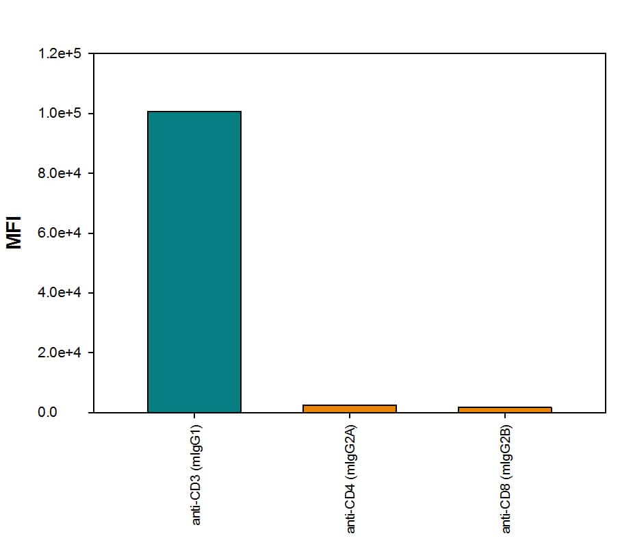
1isotype), Mouse anti-Human CD4 Monoclonal Antibody (Catalog #
MAB3791, Mouse IgG
2Aisotype), or Mouse anti-Human CD8a Monoclonal Antibody (Catalog #
MAB1509, Mouse IgG
2Bisotype), followed by Rabbit anti-Mouse IgG
1-specific secondary antibody (Catalog # MAB9627) then PE-conjugated Goat anti-Rabbit secondary antibody (Catalog #
F0110). View our protocol for
Staining Membrane-associated Proteins." class="big_lightbox">
MAB100, Mouse IgG
1isotype), Mouse anti-Human CD4 Monoclonal Antibody (Catalog #
MAB3791, Mouse IgG
2Aisotype), or Mouse anti-Human CD8a Monoclonal Antibody (Catalog #
MAB1509, Mouse IgG
2Bisotype), followed by Rabbit anti-Mouse IgG
1-specific secondary antibody (Catalog # MAB9627) then PE-conjugated Goat anti-Rabbit secondary antibody (Catalog #
F0110). View our protocol for
Staining Membrane-associated Proteins." class="big_thumb" />
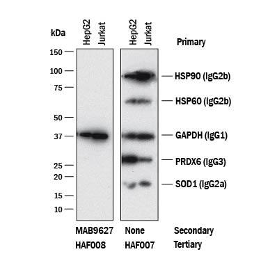
 MAB3286, Mouse IgG2Bisotype), anti-HSP60 (Catalog # MAB1800, Mouse IgG2Bisotype), anti-GAPDH (Catalog # MAB5718,Mouse IgG1isotype), anti-Peroxiredoxin 6 (Catalog # MAB3490, Mouse IgG3isotype), and anti-SOD1 (Catalog # MAB3418, Mouse IgG2Aisotype). The left panel was then probed with 0.5 µg/mL of Rabbit Anti-Mouse IgG1(Catalog # MAB9627) followed by HRP-conjugated Anti-Rabbit IgG Secondary Antibody (Catalog # HAF008). A specific band was detected for IgG1isotype antibody at approximately 39 kDa (as indicated). The right panel was probed with HRP-conjugated Anti-Mouse IgG Secondary Antibody (Catalog # HAF007) but not Rabbit Anti-Mouse IgG1Monoclonal Antibody (Catalog # MAB9627). This experiment was conducted under reducing conditions and using Immunoblot Buffer Group 1." alt="Western blot shows lysates of HepG2 and Jurkat cell lines. PVDF membrane was probed with anti-HSP90 (Catalog # MAB3286, Mouse IgG2Bisotype), anti-HSP60 (Catalog # MAB1800, Mouse IgG2Bisotype), anti-GAPDH (Catalog # MAB5718,Mouse IgG1isotype), anti-Peroxiredoxin 6 (Catalog # MAB3490, Mouse IgG3isotype), and anti-SOD1 (Catalog # MAB3418, Mouse IgG2Aisotype). The left panel was then probed with 0.5 µg/mL of Rabbit Anti-Mouse IgG1(Catalog # MAB9627) followed by HRP-conjugated Anti-Rabbit IgG Secondary Antibody (Catalog # HAF008). A specific band was detected for IgG1isotype antibody at approximately 39 kDa (as indicated). The right panel was probed with HRP-conjugated Anti-Mouse IgG Secondary Antibody (Catalog # HAF007) but not Rabbit Anti-Mouse IgG1Monoclonal Antibody (Catalog # MAB9627). This experiment was conducted under reducing conditions and using Immunoblot Buffer Group 1." class="big_thumb" />
 HAF008). A specific band was detected for IgG1at approximately 240 kDa (as indicated). Human IgG (Catalog # MAB1101) is shown as a loading control.This experiment was conducted under non-reducing conditions and using Immunoblot Buffer Group 1." alt="Western blot shows lysates of purified human IgG1, purified human IgG2(negative control), purified human IgG3(negative control), and human purified human IgG4(negative control). PVDF membrane was probed with 1 µg/mL of Rabbit Anti-Human IgG1Monoclonal Antibody (Catalog # MAB9930) followed by HRP-conjugated Anti-Rabbit IgG Secondary Antibody (Catalog # HAF008). A specific band was detected for IgG1at approximately 240 kDa (as indicated). Human IgG (Catalog # MAB1101) is shown as a loading control.This experiment was conducted under non-reducing conditions and using Immunoblot Buffer Group 1." class="big_thumb" />
 HAF018). A specific band was detected for IgG1at approximately 240 kDa (as indicated). Human IgG (Catalog # MAB1101) is shown as a loading control.This experiment was conducted under non-reducing conditions and using Immunoblot Buffer Group 1." alt="Western blot shows lysates of purified human IgG1, purified human IgG2(negative control), purified human IgG3(negative control), and human purified human IgG4(negative control). PVDF membrane was probed with 2 µg/mL of Mouse Anti-Human IgG1Monoclonal Antibody (Catalog # MAB9894) followed by HRP-conjugated Anti-Mouse IgG Secondary Antibody (Catalog # HAF018). A specific band was detected for IgG1at approximately 240 kDa (as indicated). Human IgG (Catalog # MAB1101) is shown as a loading control.This experiment was conducted under non-reducing conditions and using Immunoblot Buffer Group 1." class="big_thumb" />
 HAF018). A specific band was detected for IgG1at approximately 240 kDa (as indicated). Human IgG (Catalog # MAB1101) is shown as a loading control.This experiment was conducted under non-reducing conditions and using Immunoblot Buffer Group 1." alt="Western blot shows lysates of purified human IgG1, purified human IgG2(negative control), purified human IgG3(negative control), and human purified human IgG4(negative control). PVDF membrane was probed with 2 µg/mL of Mouse Anti-Human IgG1Monoclonal Antibody (Catalog # MAB98941) followed by HRP-conjugated Anti-Mouse IgG Secondary Antibody (Catalog # HAF018). A specific band was detected for IgG1at approximately 240 kDa (as indicated). Human IgG (Catalog # MAB1101) is shown as a loading control.This experiment was conducted under non-reducing conditions and using Immunoblot Buffer Group 1." class="big_thumb" />


![Sandwich ELISA: IgG1 Antibody (RM117) - Azide and BSA Free [NBP2-62028] - Sandwich ELISA using RM117 as the capture antibody, and NBP3-18532 as the detection antibody, followed by an AP conjugated streptavidin.](http://images.novusbio.com/fullsize/nbp2-62028_rabbit-monoclonal-igg1-antibody-rm117-sandwich-elisa-18920252340355.png "Sandwich ELISA: IgG1 Antibody (RM117) - Azide and BSA Free [NBP2-62028] - Sandwich ELISA using RM117 as the capture antibody, and NBP3-18532 as the detection antibody, followed by an AP conjugated streptavidin.")
![ELISA: IgG1 Antibody (RM117) - Azide and BSA Free [NBP2-62028] - ELISA of human immunoglobulins shows RM117 only reacts to human IgG1. No cross reactivity with Human IgG2, IgG3, IgG4, IgE, IgD, IgA, mouse IgG, rat IgG, or goat IgG. The plate was coated with 50 ng/well of different immunoglobulins. 200 ng/mL, 50 ng/mL, or 10 ng/mL of RM117 was used as the primary antibody. An alkaline phosphatase conjugated anti-rabbit IgG as the secondary antibody.](http://images.novusbio.com/fullsize/nbp2-62028_rabbit-monoclonal-igg1-antibody-rm117-elisa-189202523521111.png)
![ELISA: IgG1 Antibody (RM117) - Azide and BSA Free [NBP2-62028] - A titer ELISA using RM117. The plate was coated with different amounts of human IgG1. A serial dilution of RM117 was used as the primary antibody. An alkaline phosphatase conjugated anti-rabbit IgG as the secondary antibody.](http://images.novusbio.com/fullsize/nbp2-62028_rabbit-monoclonal-igg1-antibody-rm117-elisa-189202523474815.png)
 MAB100, Mouse IgG1isotype), Mouse anti-Human CD4 Monoclonal Antibody (Catalog #
  MAB11012) was biotinylated and incubated with the protein captured on the plate. Detection of the standard curve was achieved by incubating Streptavidin-HRP (Catalog # DY998) followed by Substrate Solution (Catalog # DY999) and stopping the enzymatic reaction with Stop Solution (Catalog # DY994)." class="big_thumb" />
 DY990). Rabbit Anti-Human IgG Monoclonal Antibody (Catalog #
 ![Western Blot: IgG1 Antibody (RM106) [Unconjugated] [NBP2-62006] - Western blot of nonreduced(-) and reduced(+) mouse IgG1, using NBP2-62006. This antibody reacts to nonreduced IgG1 (~150 kDa) stronger than the reduced g1 form (~50 kDa).](http://images.novusbio.com/fullsize/IgG1-Antibody-RM106-Unconjugated-Western-Blot-NBP2-62006-img0004.jpg "Western Blot: IgG1 Antibody (RM106) [Unconjugated] [NBP2-62006] - Western blot of nonreduced(-) and reduced(+) mouse IgG1, using NBP2-62006. This antibody reacts to nonreduced IgG1 (~150 kDa) stronger than the reduced g1 form (~50 kDa).")
![ELISA: IgG1 Antibody (RM106) - Azide and BSA Free [NBP2-62006] - A titer ELISA of mouse IgG1. The plate was coated with different amount of mouse IgG1. A serial dilution of RM106 was used as the primary antibody. An alkaline phosphatase conjugated anti-rabbit IgG as the secondary antibody.](http://images.novusbio.com/fullsize/nbp2-62006_rabbit-monoclonal-igg1-antibody-rm106-elisa-189202523521121.png)
![ELISA: IgG1 Antibody (RM106) - Azide and BSA Free [NBP2-62006] - ELISA of mouse immunoglobulins shows RM106 reacts to the Fc region of mouse IgG1; no cross reactivity with IgG2a, IgG3, IgM, IgA, IgE, human IgG, or rat IgG. The plate was coated with 50 ng/well of different immunoglobulins. 200 ng/mL, 50 ng/mL, or 10 ng/mL of RM106 was used as the primary antibody. An alkaline phosphatase conjugated anti-rabbit IgG as the secondary antibody.](http://images.novusbio.com/fullsize/nbp2-62006_rabbit-monoclonal-igg1-antibody-rm106-elisa-189202523423518.png)
![Western Blot: IgG1 Antibody (SR1920) [NBP3-22442] - Western blot analysis using NBP3-22442 in Human tonsil cell lysate.](http://images.novusbio.com/fullsize/nbp3-22442_rabbit-igg1-mab-sr1920-57202317215142.jpg "Western Blot: IgG1 Antibody (SR1920) [NBP3-22442] - Western blot analysis using NBP3-22442 in Human tonsil cell lysate.")


