DR647RB) and counterstained with DAPI (blue; Lunaphore Catalog #
DR100). Specific staining was localized to the cytoplasm and membrane. Protocol available in
COMET™ Panel Builder." class="big_lightbox">
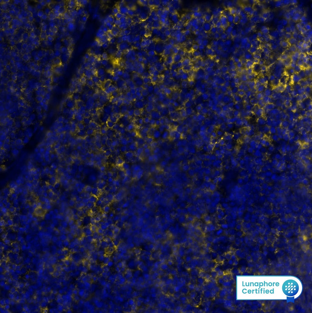
DR647RB) and counterstained with DAPI (blue; Lunaphore Catalog #
DR100). Specific staining was localized to the cytoplasm and membrane. Protocol available in
COMET™ Panel Builder." alt="CD16 was detected in perfusion fixed frozen sections of mouse Spleen using Rabbit Anti-Mouse CD16, Monoclonal Antibody (Catalog #MAB11667) at 15ug/mL at 27 ° Celsius for 4 minutes. Before incubation with the primary antibody, tissue underwent preprocessing by incubating tissue with Multi Staining Buffer (Lunaphore Catalog # BU06) for 5 minutes at room temperature followed by a 20-minute incubation inTris-Buffered Saline + 0.2% Triton at room temperature. Tissue was stained using the Alexa Fluor™ Plus 647 Goat anti-Rabbit IgG Secondary Antibody at 1:200 at 37 ° Celsius for 2 minutes. (Yellow; Lunaphore Catalog #
DR647RB) and counterstained with DAPI (blue; Lunaphore Catalog #
DR100). Specific staining was localized to the cytoplasm and membrane. Protocol available in
COMET™ Panel Builder." class="big_thumb" />
HAF008). A specific band was detected for Fc gamma RIIIB/CD16b at approximately 40-60 kDa (as indicated). This experiment was conducted under reducing conditions and using
Western Blot Buffer Group 1." class="big_lightbox">

HAF008). A specific band was detected for Fc gamma RIIIB/CD16b at approximately 40-60 kDa (as indicated). This experiment was conducted under reducing conditions and using
Western Blot Buffer Group 1." class="big_thumb" />
VC003) or the HRP-conjugated Anti-Rabbit IgG Secondary Antibody (Catalog #
HAF008). Before incubation with the primary antibody, tissue was subjected to heat-induced epitope retrieval using VisUCyte Antigen Retrieval Reagent-Basic (Catalog #
VCTS021). Tissue was stained using DAB (brown) and counterstained with hematoxylin (blue). Specific staining was localized to the cell surface of Kupffer cells. View our protocol for
IHC Staining with VisUCyte HRP Polymer Detection Reagents." class="big_lightbox">

VC003) or the HRP-conjugated Anti-Rabbit IgG Secondary Antibody (Catalog #
HAF008). Before incubation with the primary antibody, tissue was subjected to heat-induced epitope retrieval using VisUCyte Antigen Retrieval Reagent-Basic (Catalog #
VCTS021). Tissue was stained using DAB (brown) and counterstained with hematoxylin (blue). Specific staining was localized to the cell surface of Kupffer cells. View our protocol for
IHC Staining with VisUCyte HRP Polymer Detection Reagents." class="big_thumb" />
VC003) or the HRP-conjugated Anti-Rabbit IgG Secondary Antibody (Catalog #
HAF008). Before incubation with the primary antibody, tissue was subjected to heat-induced epitope retrieval using VisUCyte Antigen Retrieval Reagent-Basic (Catalog #
VCTS021). Tissue was stained using DAB (brown) and counterstained with hematoxylin (blue). Specific staining was localized to the cell surface of macrophages. View our protocol for
IHC Staining with VisUCyte HRP Polymer Detection Reagents." class="big_lightbox">

VC003) or the HRP-conjugated Anti-Rabbit IgG Secondary Antibody (Catalog #
HAF008). Before incubation with the primary antibody, tissue was subjected to heat-induced epitope retrieval using VisUCyte Antigen Retrieval Reagent-Basic (Catalog #
VCTS021). Tissue was stained using DAB (brown) and counterstained with hematoxylin (blue). Specific staining was localized to the cell surface of macrophages. View our protocol for
IHC Staining with VisUCyte HRP Polymer Detection Reagents." class="big_thumb" />
VC003) or the HRP-conjugated Anti-Rabbit IgG Secondary Antibody (Catalog #
HAF008). Before incubation with the primary antibody, tissue was subjected to heat-induced epitope retrieval using VisUCyte Antigen Retrieval Reagent-Basic (Catalog #
VCTS021). Tissue was stained using DAB (brown) and counterstained with hematoxylin (blue). Specific staining was localized to the cell surface. View our protocol for
IHC Staining with VisUCyte HRP Polymer Detection Reagents." class="big_lightbox">

VC003) or the HRP-conjugated Anti-Rabbit IgG Secondary Antibody (Catalog #
HAF008). Before incubation with the primary antibody, tissue was subjected to heat-induced epitope retrieval using VisUCyte Antigen Retrieval Reagent-Basic (Catalog #
VCTS021). Tissue was stained using DAB (brown) and counterstained with hematoxylin (blue). Specific staining was localized to the cell surface. View our protocol for
IHC Staining with VisUCyte HRP Polymer Detection Reagents." class="big_thumb" />
 DR555RT) and counterstained with DAPI (blue; Lunaphore Catalog # DR100).Specific staining was localized to the cytoplasm. Protocol available in COMET™ Panel Builder." alt="CD16b was detected in perfusion fixed paraffin-embedded sections of mouse Colon using Rat Anti-Mouse CD16b, Monoclonal Antibody (Catalog #MAB11725) at 10ug/mL at 37° Celsius for 4 minutes. Before incubation with the primary antibody, tissue underwent an all-in-one dewaxing and antigen retrieval preprocessing using PreTreatment Module (PT Module)and Dewax and HIER Buffer H (pH 9; Epredia Catalog #TA-999-DHBH). Tissue was stained using the Alexa Fluor™ 555 Goat anti-Rat IgG Secondary Antibody at 1:100 at 37 ° Celsius for 2 minutes. (Yellow; Lunaphore Catalog # DR555RT) and counterstained with DAPI (blue; Lunaphore Catalog # DR100).Specific staining was localized to the cytoplasm. Protocol available in COMET™ Panel Builder." class="big_thumb" />
 DR647RB) and counterstained with DAPI (blue; Lunaphore Catalog # DR100). Specific staining was localized to the cytoplasm and membrane. Protocol available in COMET™ Panel Builder." alt="CD16 was detected in perfusion fixed frozen sections of mouse Spleen using Rabbit Anti-Mouse CD16, Monoclonal Antibody (Catalog #MAB11667) at 15ug/mL at 27 ° Celsius for 4 minutes. Before incubation with the primary antibody, tissue underwent preprocessing by incubating tissue with Multi Staining Buffer (Lunaphore Catalog # BU06) for 5 minutes at room temperature followed by a 20-minute incubation inTris-Buffered Saline + 0.2% Triton at room temperature. Tissue was stained using the Alexa Fluor™ Plus 647 Goat anti-Rabbit IgG Secondary Antibody at 1:200 at 37 ° Celsius for 2 minutes. (Yellow; Lunaphore Catalog # DR647RB) and counterstained with DAPI (blue; Lunaphore Catalog # DR100). Specific staining was localized to the cytoplasm and membrane. Protocol available in COMET™ Panel Builder." class="big_thumb" />


 DR555RT) and counterstained with DAPI (blue; Lunaphore Catalog #
  DR555RT) and counterstained with DAPI (blue; Lunaphore Catalog #
  DR555RT) and counterstained with DAPI (blue; Lunaphore Catalog #
  HAF005). A specific band was detected for Fc gamma RIIIB/CD16b at approximately 40 kDa (as indicated). This experiment was conducted under reducing conditions and using
  VCTS021). Tissue was stained using the HRP-conjugated Anti-Rat IgG Secondary Antibody (Catalog #
  VCTS021). Tissue was stained using the HRP-conjugated Anti-Rat IgG Secondary Antibody (Catalog #
  HAF008). A specific band was detected for Fc gamma RIIIB/CD16b at approximately 40-60 kDa (as indicated). This experiment was conducted under reducing conditions and using
  VC003) or the HRP-conjugated Anti-Rabbit IgG Secondary Antibody (Catalog #
  VC003) or the HRP-conjugated Anti-Rabbit IgG Secondary Antibody (Catalog #
  VC003) or the HRP-conjugated Anti-Rabbit IgG Secondary Antibody (Catalog #
  NL004) and counterstained with DAPI (blue). Specific staining was localized to the cell surface of RAW264.7 (positive) cells. View our protocol for
 ![Western Blot: Fc gamma RIIIB/CD16b Antibody (BLR163J) [NBP3-14737] - Whole cell lysate (50 ug) from HeLa, HEK293T, PBMCs, Jurkat, and Hep-G2 cells prepared using NETN lysis buffer. Rabbit anti-CD16 recombinant monoclonal antibody used at 1:1000. HRP-conjugated goat anti-rabbit IgG. Chemiluminescence with an exposure time of 75 seconds. Lower Panel: Rabbit anti-Actin recombinant monoclonal antibody.](http://images.novusbio.com/fullsize/Fc-gamma-RIIIB-CD16b-Antibody-BLR163J-Western-Blot-NBP3-14737-img0001.jpg "Western Blot: Fc gamma RIIIB/CD16b Antibody (BLR163J) [NBP3-14737] - Whole cell lysate (50 ug) from HeLa, HEK293T, PBMCs, Jurkat, and Hep-G2 cells prepared using NETN lysis buffer. Rabbit anti-CD16 recombinant monoclonal antibody used at 1:1000. HRP-conjugated goat anti-rabbit IgG. Chemiluminescence with an exposure time of 75 seconds. Lower Panel: Rabbit anti-Actin recombinant monoclonal antibody.")
![Immunohistochemistry: Fc gamma RIIIB/CD16b Antibody (BLR163J) [NBP3-14737] - Rabbit anti-CD16 recombinant monoclonal antibody. HRP-conjugated goat anti-rabbit IgG.](http://images.novusbio.com/fullsize/Fc-gamma-RIIIB-CD16b-Antibody-BLR163J-Immunohistochemistry-NBP3-14737-img0003.jpg)
![Immunohistochemistry: Fc gamma RIIIB/CD16b Antibody (BLR163J) [NBP3-14737] - Rabbit anti-CD16 recombinant monoclonal antibody. HRP-conjugated goat anti-rabbit IgG.](http://images.novusbio.com/fullsize/Fc-gamma-RIIIB-CD16b-Antibody-BLR163J-Immunohistochemistry-NBP3-14737-img0002.jpg)
![Detection of human CD16 in FFPE CD16A transfectedHEKES cells (left) and CD16B transfected HEKES cells(right) by immunocytochemistry. Antibody: Rabbit antiCD16A recombinant monoclonal antibody [BLR163J] Secondary: HRP-conjugated goat antirabbit IgG Substrate: DAB.](http://images.novusbio.com/fullsize/nbp3-14737_rabbit-fc-gamma-riiib-cd16b-mab-immunohistochemistry-paraffin-2612026105934.png)
![Detection of human CD16 in FFPE renal cell carcinoma byimmunohistochemistry. Antibody: Rabbit anti-CD16Arecombinant monoclonal antibody [BLR163J] Secondary: HRP-conjugated goat anti-rabbit IgG Substrate: DAB.](http://images.novusbio.com/fullsize/nbp3-14737_rabbit-fc-gamma-riiib-cd16b-mab-immunohistochemistry-paraffin-26120261119.png)
![Immunohistochemistry-Paraffin: Fc gamma RIIIB/CD16b Antibody (24) [NBP3-12795] - Staining of human CD16b in human spleen with mouse monoclonal antibody at 1:2000 dilution.](http://images.novusbio.com/fullsize/Fc-gamma-RIIIB-CD16b-Antibody-24-Immunohistochemistry-Paraffin-NBP3-12795-img0002.jpg "Immunohistochemistry-Paraffin: Fc gamma RIIIB/CD16b Antibody (24) [NBP3-12795] - Staining of human CD16b in human spleen with mouse monoclonal antibody at 1:2000 dilution.")
![Immunohistochemistry-Paraffin: Fc gamma RIIIB/CD16b Antibody (24) [NBP3-12795] - Staining of human CD16b in human liver with mouse monoclonal antibody at 1:2000 dilution.](http://images.novusbio.com/fullsize/Fc-gamma-RIIIB-CD16b-Antibody-24-Immunohistochemistry-Paraffin-NBP3-12795-img0003.jpg)
![Immunohistochemistry-Paraffin: Fc gamma RIIIB/CD16b Antibody [NBP3-41105] - DAB staining on IHC-P; Samples: Human Small intestine Tissue; Primary Ab: 20ug/ml NBP3-41105; Second Ab: 2ug/mL HRP-Linked Caprine Anti-Rabbit IgG Polyclonal Antibody](http://images.novusbio.com/fullsize/nbp3-41105_rabbit-fc-gamma-riiib-cd16b-pab-612202421262929.jpg "Immunohistochemistry-Paraffin: Fc gamma RIIIB/CD16b Antibody [NBP3-41105] - DAB staining on IHC-P; Samples: Human Small intestine Tissue; Primary Ab: 20ug/ml NBP3-41105; Second Ab: 2ug/mL HRP-Linked Caprine Anti-Rabbit IgG Polyclonal Antibody")
![Western Blot: Fc gamma RIIIB/CD16b Antibody [NBP3-41105] - Sample: Recombinant protein.](http://images.novusbio.com/fullsize/nbp3-41105_rabbit-fc-gamma-riiib-cd16b-pab-612202421373057.jpg)
![Immunocytochemistry /Immunofluorescence: Fc gamma RIIIB/CD16b Antibody (C1) [NBP3-41587] - FITC staining on IF; Sample: Human U2OS cell; Primary Ab: 30ug/ml NBP3-41587; Second Ab: 5ug/ml FITC-Linked Caprine Anti-Mouse IgG Polyclonal Antibody](http://images.novusbio.com/fullsize/nbp3-41587_mouse-fc-gamma-riiib-cd16b-mab-c1-612202421121382.jpg "Immunocytochemistry /Immunofluorescence: Fc gamma RIIIB/CD16b Antibody (C1) [NBP3-41587] - FITC staining on IF; Sample: Human U2OS cell; Primary Ab: 30ug/ml NBP3-41587; Second Ab: 5ug/ml FITC-Linked Caprine Anti-Mouse IgG Polyclonal Antibody")


