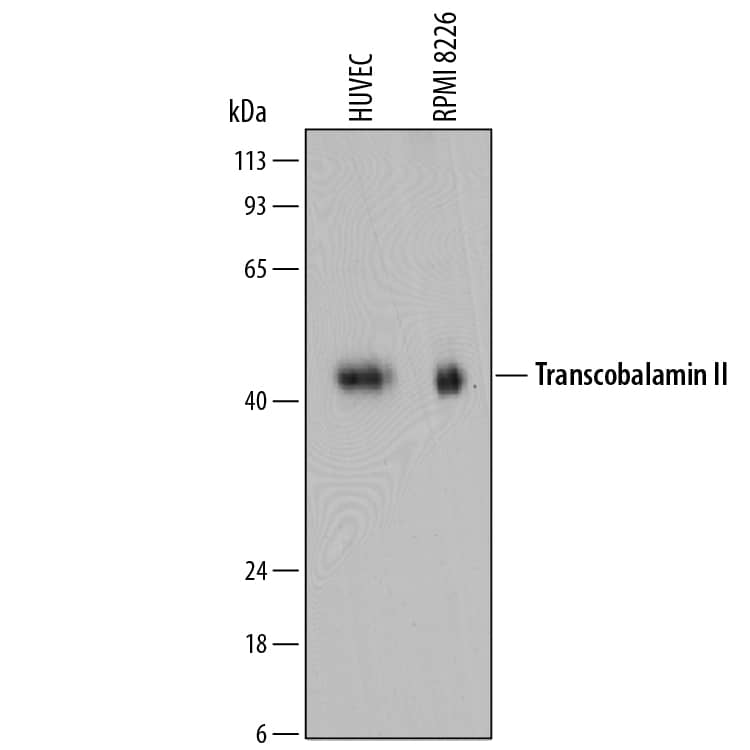
 HAF016). A specific band was detected for Transcobalamin II at approximately 43 kDa (as indicated). This experiment was conducted under reducing conditions and using Immunoblot Buffer Group 1." alt="Western blot shows lysates of HUVEC human umbilical vein endothelial cells and RPMI 8226 human multiple myeloma cell line. PVDF membrane was probed with 1 µg/mL of Sheep Anti-Human/Mouse/Rat Transcobalamin II Antigen Affinity-purified Polyclonal Antibody (Catalog # AF7895) followed by HRP-conjugated Anti-Sheep IgG Secondary Antibody (Catalog # HAF016). A specific band was detected for Transcobalamin II at approximately 43 kDa (as indicated). This experiment was conducted under reducing conditions and using Immunoblot Buffer Group 1." class="big_thumb" />
| Formulation | Catalog # | Availability | Price | |
|---|---|---|---|---|
| Carrier Free | AF7895-SP | |||
 HAF018). A specific band was detected for Transcobalamin II at approximately 43 kDa (as indicated). This experiment was conducted under reducing conditions and using Immunoblot Buffer Group 1." alt="Western blot shows lysates of HUVEC human umbilical vein endothelial cells. PVDF membrane was probed with 2 µg/mL of Mouse Anti-Human Transcobalamin II Monoclonal Antibody (Catalog # MAB7895) followed by HRP-conjugated Anti-Mouse IgG Secondary Antibody (Catalog # HAF018). A specific band was detected for Transcobalamin II at approximately 43 kDa (as indicated). This experiment was conducted under reducing conditions and using Immunoblot Buffer Group 1." class="big_thumb" />
| Formulation | Catalog # | Availability | Price | |
|---|---|---|---|---|
| Carrier Free | MAB7895-SP | |||
| Formulation | Catalog # | Availability | Price | |
|---|---|---|---|---|
| BSA Free | NBP2-94355-0.02ml | |||


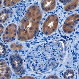 CTS013). Tissue was stained using the Anti-Sheep HRP-DAB Cell & Tissue Staining Kit (brown; Catalog #
![Western Blot: Transcobalamin II Antibody (2F4) [H00006948-M01] - Analysis of TCN2 expression in transfected 293T cell line by TCN2 monoclonal antibody (M01), clone 2F4.Lane 1: TCN2 transfected lysate (Predicted MW: 47.5 KDa).Lane 2: Non-transfected lysate.](http://images.novusbio.com/fullsize/Transcobalamin-II-Antibody-2F4-Western-Blot-H00006948-M01-img0002.jpg "Western Blot: Transcobalamin II Antibody (2F4) [H00006948-M01] - Analysis of TCN2 expression in transfected 293T cell line by TCN2 monoclonal antibody (M01), clone 2F4.Lane 1: TCN2 transfected lysate (Predicted MW: 47.5 KDa).Lane 2: Non-transfected lysate.")
![Sandwich ELISA: Transcobalamin II Antibody (2F4) [H00006948-M01] - Detection limit for recombinant GST tagged TCN2 is 1 ng/ml as a capture antibody.](http://images.novusbio.com/fullsize/Transcobalamin-II-Antibody-2F4-Sandwich-ELISA-H00006948-M01-img0001.jpg)
![Western Blot: Transcobalamin II Antibody [NBP2-94355] - Analysis of extracts of various cell lines, using Transcobalamin II at 1:1000 dilution. Secondary antibody: HRP Goat Anti-Rabbit IgG (H+L) at 1:10000 dilution. Lysates/proteins: 25ug per lane. Blocking buffer: 3% nonfat dry milk in TBST. Detection: ECL Basic Kit . Exposure time: 30s.](http://images.novusbio.com/fullsize/Transcobalamin-II-Antibody-Western-Blot-NBP2-94355-img0002.jpg "Western Blot: Transcobalamin II Antibody [NBP2-94355] - Analysis of extracts of various cell lines, using Transcobalamin II at 1:1000 dilution. Secondary antibody: HRP Goat Anti-Rabbit IgG (H+L) at 1:10000 dilution. Lysates/proteins: 25ug per lane. Blocking buffer: 3% nonfat dry milk in TBST. Detection: ECL Basic Kit . Exposure time: 30s.")
![Western Blot: Transcobalamin II Antibody [NBP1-74220] - Mouse Pancreas Lysate 1.0 ug/ml, gel concentration 12%](http://images.novusbio.com/fullsize/Transcobalamin-II-Antibody-Western-Blot-NBP1-74220-img0002.jpg "Western Blot: Transcobalamin II Antibody [NBP1-74220] - Mouse Pancreas Lysate 1.0 ug/ml, gel concentration 12%")
![Western Blot: TCN2 Overexpression Lysate (Adult Normal) [NBP2-10251] Left-Empty vector transfected control cell lysate (HEK293 cell lysate); Right -Over-expression Lysate for TCN2.](http://images.novusbio.com/fullsize/TCN2-Overexpression-Lysate-Adult-Normal-Western-Blot-NBP2-10251-img0001.jpg "Western Blot: TCN2 Overexpression Lysate (Adult Normal) [NBP2-10251] Left-Empty vector transfected control cell lysate (HEK293 cell lysate); Right -Over-expression Lysate for TCN2.")
![Western Blot: TCN2 Overexpression Lysate (Adult Normal) [NBL1-16784] Left-Empty vector transfected control cell lysate (HEK293 cell lysate); Right -Over-expression Lysate for TCN2.](http://images.novusbio.com/fullsize/TCN2-Overexpression-Lysate-Adult-Normal-Western-Blot-NBL1-16784-img0002.jpg "Western Blot: TCN2 Overexpression Lysate (Adult Normal) [NBL1-16784] Left-Empty vector transfected control cell lysate (HEK293 cell lysate); Right -Over-expression Lysate for TCN2.")
![ELISA: Human Transcobalamin II ELISA Kit (Colorimetric) [NBP3-38684] - Standard Curve Reference](http://images.novusbio.com/fullsize/nbp3-38684_human-transcobalamin-ii-elisa-kit-colorimetric-211020241538032.png "ELISA: Human Transcobalamin II ELISA Kit (Colorimetric) [NBP3-38684] - Standard Curve Reference")
![ELISA: Human Transcobalamin II - Ready-To-Use ELISA Kit (Colorimetric) [NBP3-38683] - Standard Curve Reference](http://images.novusbio.com/fullsize/nbp3-38683_human-transcobalamin-ii-ready-to-use-elisa-kit-color-2110202415355074.png "ELISA: Human Transcobalamin II - Ready-To-Use ELISA Kit (Colorimetric) [NBP3-38683] - Standard Curve Reference")


