| Immunogen | A synthetic peptide (RSFADRCKEVQQI) corresponding to the N-terminal of human MAP1LC3 A protein conjugated to Blue Carrier Protein has been used as the immunogen. |
| Localization | Accession Number: MLP3A_HUMAN ; MLP3A_MOUSE ; MLP3A_RAT |
| Marker | Autophagosome Marker |
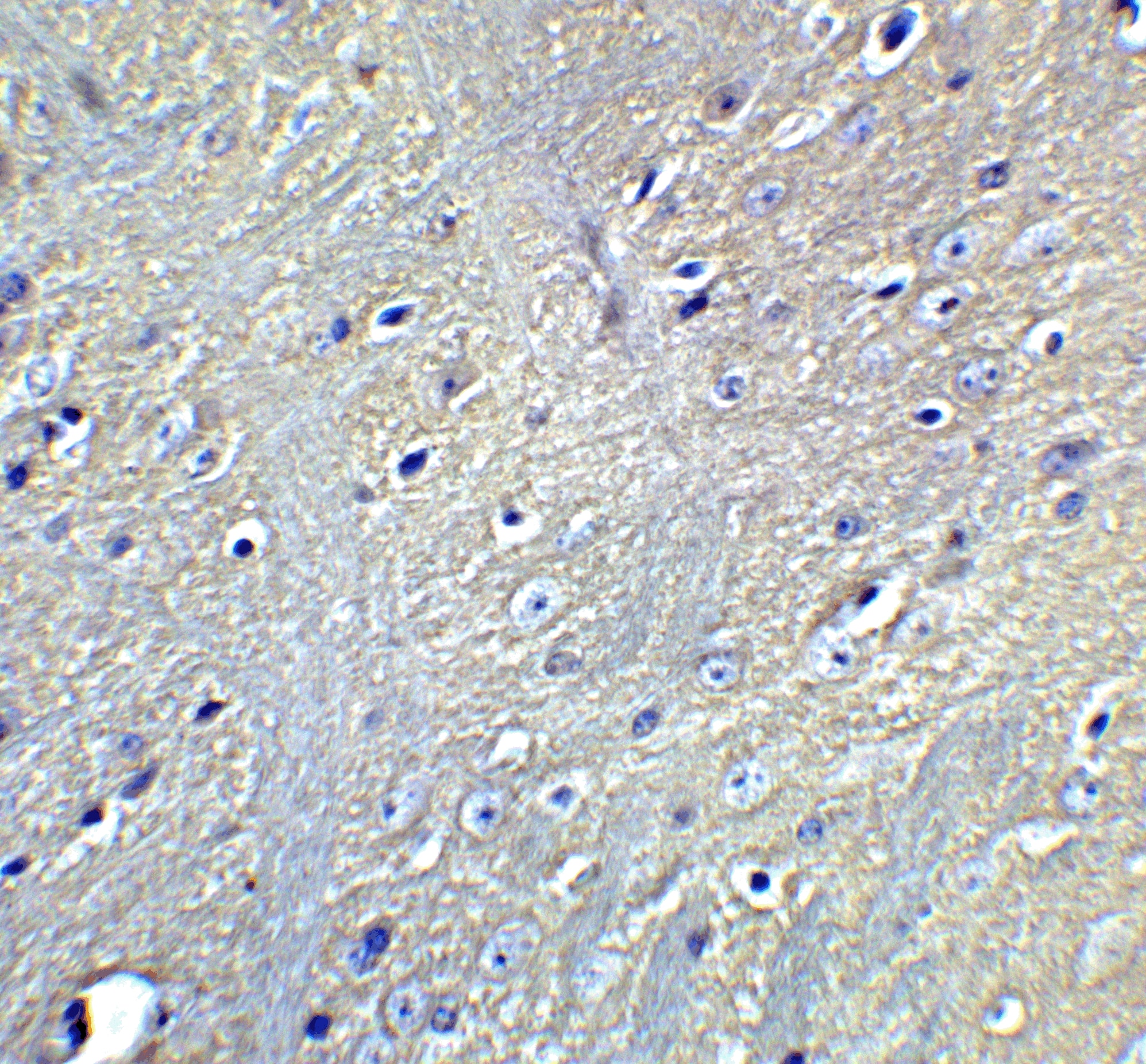
| Specificity | IHC, WB and ELISA confirmed the specificity for MAP1LC3 A (MAP1LC3A, APG8a). |
| Clonality | Polyclonal |
| Host | Rabbit |
| Gene | MAP1LC3A |
| Purity | Unpurified |
| Innovator's Reward | Test in a species/application not listed above to receive a full credit towards a future purchase. |
| Dilutions |
|
|
| Application Notes | Immunofluorescence and WB. A dilution of 1:100 to 1:1000 dilution is recommended for these applications. The optimal dilution should be determined by the end user. Although not tested this antibody may be useful in IHC-P. |
|
| Publications |
|
| Storage | Store at 4C short term. Aliquot and store at -20C long term. Avoid freeze-thaw cycles. |
| Buffer | Whole antisera |
| Preservative | No Preservative |
| Concentration | LYOPH |
| Purity | Unpurified |
| Reconstitution Instructions | Reconstitute with deionized water. Reconstitute with 0.1 ml sterilized water. Centrifuge to remove any insoluble material. |


![Western Blot AKT [p Ser473] Antibody [Unconjugated] - Pan Specific](https://images.novusbio.com/images2/Akt3_AF887_Western_Blot_5930.jpg)
![Simple Western AKT [p Ser473] Antibody [Unconjugated] - Pan Specific](https://images.novusbio.com/images2/16332.jpg)
![Intracellular Staining by Flow Cytometry AKT [p Ser473] Antibody [Unconjugated] - Pan Specific](https://images.novusbio.com/images2/Akt3_AF887_Flow_Cytometry_8283.jpg)
![Immunohistochemistry ATG7 Antibody (683906) [Unconjugated]](https://images.novusbio.com/images2/ATG7_MAB6608_Immunohistochemistry_10631.jpg)
![Simple Western ATG7 Antibody (683906) [Unconjugated]](https://images.novusbio.com/images2/ATG7_MAB6608_Simple_Western_16409.jpg)
![Western Blot ATG7 Antibody (683906) [Unconjugated]](https://images.novusbio.com/images2/ATG7_MAB6608_Western_Blot_11614.jpg)


| Publication using R-146-100 | Applications | Species |
|---|---|---|
| The MGC Project Team. Genome Res. 14:2121-2127. 2004-01-01 [PMID: 15489334] |
Secondary Antibodies |
Isotype Controls |
Research Areas for LC3A Antibody (R-146-100)Find related products by research area.
|
|
Read full blog post. |
|
Losing memory: Toxicity from mutant APP and amyloid beta explain the hippocampal neuronal damage in Alzheimer's disease By Jamshed Arslan Pharm.D. Alzheimer's disease (AD) is an irreversible brain disorder that destroys memory and thinking skills. The telltale signs of AD brains are extracellular deposits of amy... Read full blog post. |
|
Nuclear LC3: Why is it there and what is it doing? By Christina Towers, PhD. Cells use the complex process of autophagy to degrade and recycle cytoplasmic material. There are over 20 proteins that have been implicated in this process and appropriately named core ... Read full blog post. |
|
Why LC3B Antibodies Make Ideal Autophagosomes Membrane Markers The human form of microtubule-associated protein light chain 3 (LC3) is expressed as 3 splice variants LC3A, LC3B, and LC3C.1 LC3B is a subunit of the MAP1A and MAP1B microtubule-binding proteins and plays a central role in autophagosome membrane stru... Read full blog post. |
The concentration calculator allows you to quickly calculate the volume, mass or concentration of your vial. Simply enter your mass, volume, or concentration values for your reagent and the calculator will determine the rest.


![Immunohistochemistry-Paraffin TOR/mTOR [p Ser2448] Antibody - BSA Free](https://images.novusbio.com/images/TOR-mTOR-[p-Ser2448]-Antibody-Immunohistochemistry-Paraffin-NB600-607-img0005.jpg)
![Western Blot TOR/mTOR [p Ser2448] Antibody - BSA Free](https://images.novusbio.com/images/TOR-mTOR-[p-Ser2448]-Antibody-Western-Blot-NB600-607-img0006.jpg)
![Data TOR/mTOR [p Ser2448] Antibody - BSA Free](https://images.novusbio.com/images/TOR-mTOR-[p-Ser2448]-Antibody-N-A-NB600-607-img0008.jpg)
![Immunocytochemistry Caspase-3 Antibody [Unconjugated] - Active](https://images.novusbio.com/images2/Caspase-3_AF835_Immunocytochemistry_6532.jpg)
![Immunohistochemistry Caspase-3 Antibody [Unconjugated] - Active](https://images.novusbio.com/images2/Caspase3_AF835_Immunohistochemistry_22976.jpg)
![Immunocytochemistry Caspase-3 Antibody [Unconjugated] - Active](https://images.novusbio.com/images2/Caspase-3_AF835_Immunocytochemistry_9340.jpg)


![Western Blot ERK2 Antibody [Unconjugated]](https://images.novusbio.com/images2/ERK2_AF1230_Western_Blot_5097.jpg)
![Immunohistochemistry ERK2 Antibody [Unconjugated]](https://images.novusbio.com/images2/ERK2_AF1230_Immunohistochemistry_20696.jpg)
![Knockout Validated ERK2 Antibody [Unconjugated]](https://images.novusbio.com/images2/ERK2_AF1230_Knockout_Validated_22864.jpg)


