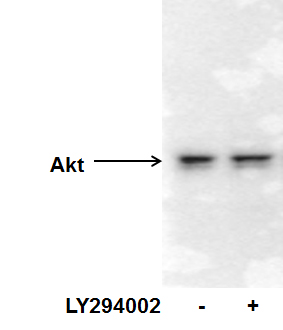
| Immunogen | Synthetic peptide corresponding to amino acids 460-480 of the C-terminal of human, rat, mouse and chicken AKT protein conjugated to KLH. |
| Localization | Cytoplasmic, and nuclear after activation by integrin linked protein kinase 1 (ILK1). |
| Specificity | AKT1 |
| Clonality | Polyclonal |
| Host | Rabbit |
| Gene | AKT1 |
| Purity | Unpurified |
| Innovator's Reward | Test in a species/application not listed above to receive a full credit towards a future purchase. |
| Dilutions |
|
||
| Theoretical MW | 55.7 kDa. Disclaimer note: The observed molecular weight of the protein may vary from the listed predicted molecular weight due to post translational modifications, post translation cleavages, relative charges, and other experimental factors. |
||
| Control |
|
||
| Reviewed Applications |
|
| Storage | Aliquot and store at -20C or -80C. Avoid freeze-thaw cycles. |
| Buffer | 0.02 M Potassium Phosphate, 0.15 M Sodium Chloride, pH 7.2 |
| Preservative | 0.01% Sodium Azide |
| Purity | Unpurified |
![Simple Western EphB2 Antibody [Unconjugated]](https://images.novusbio.com/images2/EphB2_AF467_Simple_Western_21454.jpg)
![Western Blot EphB2 Antibody [Unconjugated]](https://images.novusbio.com/images2/EphB2_AF467_Western_Blot_21511.jpg)
![Immunocytochemistry EphB2 Antibody [Unconjugated]](https://images.novusbio.com/images2/EphB2_AF467_Immunocytochemistry__Immunofluorescence_17288.jpg)
![Immunohistochemistry EGFR Antibody [Unconjugated]](https://images.novusbio.com/images2/EGF_R_AF231_Immunohistochemistry_20892.jpg)
![Immunocytochemistry EGFR Antibody [Unconjugated]](https://images.novusbio.com/images2/EGF_R_AF231_Immunocytochemistry__Immunofluorescence_21143.jpg)
![Western Blot EGFR Antibody [Unconjugated]](https://images.novusbio.com/images2/EGF_R_AF231_Western_Blot_19925.jpg)
![Western Blot PTEN Antibody [Unconjugated]](https://images.novusbio.com/images2/PTEN_AF847_Western_Blot_5922.jpg)
![Simple Western PTEN Antibody [Unconjugated]](https://images.novusbio.com/images2/16342.jpg)
![Knockout Validated PTEN Antibody [Unconjugated]](https://images.novusbio.com/images2/PTEN_AF847_Knockout_Validated_22996.jpg)


| Images | Ratings | Applications | Species | Date | Details | ||||||
|---|---|---|---|---|---|---|---|---|---|---|---|
Enlarge |
reviewed by:
Verified Customer |
WB | Human | 09/08/2015 |
Summary
|
||||||
Enlarge |
reviewed by:
Hongxia Hu |
WB | Human | 09/12/2012 |
Summary
|
||||||
|
reviewed by:
Verified Customer |
WB | Mouse | 11/09/2011 |
Summary
|
Secondary Antibodies |
Isotype Controls |
Research Areas for AKT1 Antibody (NB600-467)Find related products by research area.
|
|
Understanding ‘Y’ in Breast Cancer: Crucial Role of DNA/RNA-binding Protein YB-1 in the Development, Pre-Invasive, and Metastatic Phases Jamshed Arslan, Pharm D, PhD In the United States, 1 in 8 women will be diagnosed with breast cancer in her lifetime.1 Despite the prevalence, cancer genesis is a mystery. The heterogeneity of cancers makes it diff... Read full blog post. |
|
Tips to Optimize Your Western Blot for Phosphorylated Protein Detection By Jamshed Arslan, Pharm. D., PhD. Protein phosphorylation refers to a reversible post-translational modification in which a protein kinase adds a phosphate group to an amino acid residue of a target protein. Protein ... Read full blog post. |
|
Sample collection from mammalian culture cells for kinomic analysis By Jamshed Arslan Pharm.D., PhD.IntroductionKinome describes kinases, and kinomics refers to the kinase signaling. Studying the effects of reagent (exogenously applied growth factor or inhibitor) on kinase activit... Read full blog post. |
|
Using a STAT3 antibody in chromatin immunoprecipitation (ChIP) Signal transducer and activator of transcription 3 (STAT3) is an important oncogenic transcriptional factor that mediates tumor induced immune suppression. Specifically, STAT3 transmits signals from cytokines and growth factor receptors in the pla... Read full blog post. |
|
Altered expression of BCL2 in cancer Similar to other cell processes, the balance between cell survival and cell death is an important equilibrium that when altered expression of genes can lead to a variety of disease. For example, too little cell death can promote cell overgrowth a... Read full blog post. |
|
AKT1 - Regulating cell growth and survival through phosphorylation AKT1 is a serine/threonine protein kinase with homology to protein kinase A (PKA) and protein kinase C (PKC). AKT1 contains the central kinase domain sandwiched between a pleckstrin homology domain and a regulatory domain (1). AKT1 is regulated by ... Read full blog post. |
|
Akt1 - a central player in cell survival signaling Akt1 is one of three isoforms of Akt belonging to the AGC family of serine/threonine kinases (Akt1, Akt2, and Akt3). All Akt isoforms contain an N-terminal Plekstrin Homology (PH) domain, a C-terminal regulatory domain, and a central catalytic kin... Read full blog post. |
|
AKT1, Scene 1: The Cell Must Go On Akt1 is a serine/threonine-specific protein kinase involved in many cellular signaling pathways. The major function of this kinase is to mediate cell survival, but it also plays key roles in various other cellular functions such as glycogen synthesis ... Read full blog post. |
|
Caspase 9 and Mitochondrial Apoptosis Regulation Caspase 9 (also termed ICE-LAP6, Mch6, Apaf-3) is a member of cysteine protease family of caspases and is encoded by the CASP9 gene in humans. Caspase-9 is involved in mitochondrial apoptosis pathway and is an initiator caspase. Pro-caspase-9 is activ... Read full blog post. |
|
Mapping Signal Transduction with mTOR Antibodies The protein encoded by mTOR (mammalian target of rapamycin), also known as dTOR in Drosophila, belongs to a family of phosphatidylinositol kinase-related kinases. These kinases regulate fundamental processes of cell growth, proliferation, metabolism... Read full blog post. |
The concentration calculator allows you to quickly calculate the volume, mass or concentration of your vial. Simply enter your mass, volume, or concentration values for your reagent and the calculator will determine the rest.
5 | |
4 | |
3 | |
2 | |
1 |
| Verified Customer 09/08/2015 |
||
| Application: | WB | |
| Species: | Human |
| Hongxia Hu 09/12/2012 |
||
| Application: | WB | |
| Species: | Human |
| Verified Customer 11/09/2011 |
||
| Application: | WB | |
| Species: | Mouse |


![Western Blot ERK2 Antibody [Unconjugated]](https://images.novusbio.com/images2/ERK2_AF1230_Western_Blot_5097.jpg)
![Immunohistochemistry ERK2 Antibody [Unconjugated]](https://images.novusbio.com/images2/ERK2_AF1230_Immunohistochemistry_20696.jpg)
![Knockout Validated ERK2 Antibody [Unconjugated]](https://images.novusbio.com/images2/ERK2_AF1230_Knockout_Validated_22864.jpg)
![N/A ERK1 [p Thr202, p Tyr204] [Biotin]](https://images.novusbio.com/images2/dyc1825-2_phospho-erk1-duoset-ic-economy-pack-15-plate-17122020121349.png)
![N/A ERK1 [p Thr202, p Tyr204] [Biotin]](https://images.novusbio.com/images2/dyc1825-2_phospho-erk1-duoset-ic-economy-pack-15-plate-17122020122737.png)
![N/A ERK1 [p Thr202, p Tyr204] [Biotin]](https://images.novusbio.com/images2/dyc1825-2_phospho-erk1-duoset-ic-economy-pack-15-plate-17122020122919.png)
![Simple Western PYK2/FAK2 [p Tyr402] Antibody (592918) [Unconjugated]](https://images.novusbio.com/images2/PYK2_MAB6210_Simple_Western_16639.jpg)
![Western Blot PYK2/FAK2 [p Tyr402] Antibody (592918) [Unconjugated]](https://images.novusbio.com/images2/PYK2_MAB6210_Western_Blot_9430.jpg)
![Western Blot PYK2/FAK2 [p Tyr402] Antibody (592918) [Unconjugated]](https://images.novusbio.com/images/mab6210_human-phospho-pyk2-y402-mab-clone-592918-western-blot-1212202585785.jpg)
![Immunohistochemistry-Paraffin TOR/mTOR [p Ser2448] Antibody - BSA Free](https://images.novusbio.com/images/TOR-mTOR-[p-Ser2448]-Antibody-Immunohistochemistry-Paraffin-NB600-607-img0005.jpg)
![Western Blot TOR/mTOR [p Ser2448] Antibody - BSA Free](https://images.novusbio.com/images/TOR-mTOR-[p-Ser2448]-Antibody-Western-Blot-NB600-607-img0006.jpg)
![Data TOR/mTOR [p Ser2448] Antibody - BSA Free](https://images.novusbio.com/images/TOR-mTOR-[p-Ser2448]-Antibody-N-A-NB600-607-img0008.jpg)
![Immunocytochemistry Insulin Antibody (182410) [Unconjugated]](https://images.novusbio.com/images2/Insulin_MAB1417_Immunocytochemistry_9376.jpg)
![Immunohistochemistry Insulin Antibody (182410) [Unconjugated]](https://images.novusbio.com/images2/mab1417_human-bovine-mouse-insulin-mab-clone-182410-immunohistochemistry-308202115145.jpg)
![SDS-PAGE TNF-alpha [Unconjugated]](https://images.novusbio.com/images2/TNF-alpha_210-TA_256.jpg)
![Bioactivity TNF-alpha [Unconjugated]](https://images.novusbio.com/images2/TNFalpha_210TA_1658.jpg)
![SEC-MALS TNF-alpha [Unconjugated]](https://images.novusbio.com/images/210-ta_recombinant-human-tnf-alpha-protein-sec-mals-35202312244..jpg)
![N/A VEGF [HRP]](https://images.novusbio.com/images2/DATA_VEGF_DVE00_ELISA_871.jpg)
![N/A VEGF [HRP]](https://images.novusbio.com/images2/DATA_VEGF_DVE00_ELISA_872.jpg)
![N/A VEGF [HRP]](https://images.novusbio.com/images2/VEGF_DVE00_ELISA_208.jpg)
![Immunocytochemistry Caspase-3 Antibody [Unconjugated] - Active](https://images.novusbio.com/images2/Caspase-3_AF835_Immunocytochemistry_6532.jpg)
![Immunohistochemistry Caspase-3 Antibody [Unconjugated] - Active](https://images.novusbio.com/images2/Caspase3_AF835_Immunohistochemistry_22976.jpg)
![Immunocytochemistry Caspase-3 Antibody [Unconjugated] - Active](https://images.novusbio.com/images2/Caspase-3_AF835_Immunocytochemistry_9340.jpg)
![SDS-PAGE IGF-I/IGF-1 [Unconjugated]](https://images.novusbio.com/images2/IGF-I_291-G1_40.jpg)
![Bioactivity IGF-I/IGF-1 [Unconjugated]](https://images.novusbio.com/images2/IGF-I_291-G1_41.jpg)
![Mass Spectrometry IGF-I/IGF-1 [Unconjugated]](https://images.novusbio.com/images2/IGF-I_291-G1_42.jpg)
![Western Blot JNK1 Antibody (228601) [Unconjugated]](https://images.novusbio.com/images2/JNK1_MAB17761_Western_Blot_5991.jpg)
![Western Blot JNK1 Antibody (228601) [Unconjugated]](https://images.novusbio.com/images2/JNK1_MAB17761_Western_Blot_6327.jpg)
![Immunocytochemistry JNK1 Antibody (228601) [Unconjugated]](https://images.novusbio.com/images2/JNK1_MAB17761_Immunocytochemistry__Immunofluorescence_20184.jpg)

